Hello! Welcome to your next lesson in the "Foundations in Clinical Cardiology and Diagnostics" module.
In our previous lesson, we explored how cardiac biomarkers like troponins and natriuretic peptides help us diagnose myocardial injury and hemodynamic stress. Today, we shift our focus from detecting the consequences of ischemia to proactively identifying its presence using functional testing.
This lesson will help you master the learning outcome: Interpret the results and clinical implications of exercise stress testing and myocardial perfusion imaging.
We will cover the two main pillars of functional testing for coronary artery disease (CAD):
- Exercise Stress Testing (EST), commonly known as the Treadmill Test (TMT), which uses ECG changes and physiological responses to detect ischemia.
- Myocardial Perfusion Imaging (MPI), a nuclear medicine technique that provides direct images of blood flow to the heart muscle, often in conjunction with a stress test.
You will learn the principles behind each test, how to interpret their findings, and, most importantly, how to use their results to assess a patient's risk and guide clinical decisions. This is a core topic for both clinical practice and your postgraduate entrance exams.
1. The Exercise Stress Test (EST / TMT)
The exercise stress test is often the first-line investigation for patients with suspected CAD who are able to exercise and have a normal, interpretable baseline ECG. It's a non-invasive, widely available, and cost-effective tool.
The Principle and Procedure
The goal of an EST is to increase myocardial oxygen demand through exercise and observe for evidence of ischemia, which manifests as ECG changes, symptoms, or abnormal hemodynamic responses.
To begin, let's watch a short video, 'Exercise Treadmill Test TMT', which provides an excellent overview of the procedure and the most common protocol used.
Watch the first two minutes of the video (00:00 - 02:04). Focus on understanding: The purpose of the test. The structure of the standard Bruce protocol, noting how the speed and incline increase at each stage.
A key objective during the test is for the patient to reach at least 85% of their age-predicted maximum heart rate (MPHR), calculated as . If a test is stopped before reaching this target and no ischemic changes are seen, it's considered nondiagnostic because the heart wasn't stressed enough to reliably provoke ischemia.
Interpreting the Results
A comprehensive interpretation of an EST involves more than just looking at the ECG. You must synthesize four key pieces of information.
1. ECG Changes: The Core of Diagnosis
The most important diagnostic finding is ST-segment depression.
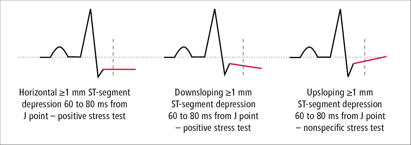
- Positive for Ischemia: ≥ 1 mm of horizontal or downsloping ST-segment depression, measured 60-80 milliseconds after the J-point.
- Non-specific: Upsloping ST-segment depression is less specific for ischemia.
- ST-segment elevation (in leads without pre-existing Q waves) is a rare but ominous sign, suggesting severe, transmural ischemia.
Stress testing and noninvasive coronary imaging
Let's reinforce these ECG criteria by reading a concise summary. This article, 'Stress testing and noninvasive coronary imaging', provides a clear description.
Read the subsections 'Test features' and 'Diagnostic and prognostic features' under the main heading 'ELECTROCARDIOGRAPHY EXERCISE STRESS TESTING'. Pay special attention to the description of a positive test and the conditions that make an ECG uninterpretable.
2. Functional Capacity: A Powerful Prognostic Indicator
How much work a patient can do is one of the most powerful predictors of their long-term survival. This is measured in Metabolic Equivalents (METs).
- 1 MET = Oxygen consumption at rest.
- < 5 METs = Poor prognosis.
-
10 METs = Excellent prognosis, regardless of ECG changes.
3. Hemodynamic Response
- Blood Pressure: Systolic blood pressure (SBP) should rise with exercise. A drop in SBP > 10 mmHg (exercise-induced hypotension) is a high-risk feature, suggesting severe multivessel disease or left ventricular dysfunction.
- Heart Rate: Failure to achieve 85% of MPHR (chronotropic incompetence) or slow heart rate recovery after exercise are also poor prognostic signs.
4. Symptoms
The development of typical angina during the test, especially when accompanied by ECG changes, increases the likelihood of significant CAD.
Indications, Contraindications, and Limitations
It's crucial to know when to order an EST and when to choose a different test.
Overview of Exercise Stress Testing - PMC
This article, 'Overview of Exercise Stress Testing', contains an excellent table summarizing the key indications and contraindications.
Please review 'Table 1. Common indications and contraindications for exercise stress testing'. This is a high-yield table for exams.
Key limitations to remember:
- The test is uninterpretable in patients with baseline ECG abnormalities like LBBB, ventricular paced rhythm, or significant resting ST depression.
- The patient must be able to exercise adequately.
- It has lower diagnostic accuracy compared to imaging tests, with a notable rate of false positives, especially in younger women.
Test your understanding!
A 55-year-old man undergoes an EST for atypical chest pain. He exercises for 11 minutes on the Bruce protocol (>10 METs) and achieves 95% of his MPHR. At peak exercise, he develops 2 mm of horizontal ST depression in leads II, III, and aVF but remains asymptomatic. How would you interpret the overall result of this test?
Show answer
This is an electrically positive stress test, indicating myocardial ischemia. However, the patient's excellent functional capacity (>10 METs) confers a very good prognosis. This is a classic example of discordant findings. While the ECG suggests ischemia, his excellent exercise tolerance suggests he is at low risk for a major cardiac event in the near future. This information is crucial for counseling the patient and deciding on the next steps, which might involve medical management and risk factor control rather than immediate invasive investigation.
2. Myocardial Perfusion Imaging (MPI)
When a standard EST is not an option or when greater diagnostic certainty is needed, we turn to MPI. This test adds a layer of imaging to the stress test, allowing us to directly see blood flow to the heart muscle.
The Principle: The Ischemic Cascade
MPI is more sensitive than EST because it detects an earlier step in the process of ischemia.
Stress Nuclear: Methodology and Case Studies (Faisal Nabi, MD) January 28, 2020
This video, presented by Dr. Faisal Nabi, provides a comprehensive look at nuclear stress testing. Let's start with the fundamental concept that makes MPI so powerful: the ischemic cascade.
Watch the segment from 00:45 to 02:45. Focus on the sequence of events in the ischemic cascade and where perfusion abnormalities fit in.
As the video explains, the ischemic cascade follows this order:
- Perfusion Abnormality (detected by MPI)
- Diastolic Dysfunction
- Systolic Dysfunction / Wall Motion Abnormality (detected by Stress Echo)
- ECG Changes (detected by EST)
- Chest Pain (Angina)
How MPI is Performed
An MPI study has two key components: the stressor and the radiotracer.
1. The Stressor: Exercise vs. Pharmacologic
Just like with EST, exercise on a treadmill is the preferred stressor because it provides valuable prognostic information about functional capacity.
For patients who cannot exercise, we use pharmacologic stress agents:
- Vasodilators (Regadenoson, Adenosine): These are the most common. They dramatically increase blood flow in normal coronary arteries but have little effect on arteries narrowed by stenosis. This creates a flow disparity ("coronary steal") that the scan can detect.
- Inotropes (Dobutamine): This agent increases heart rate and contractility, mimicking the oxygen demand of exercise.
Stress Nuclear: Methodology and Case Studies (Faisal Nabi, MD) January 28, 2020
Let's return to Dr. Nabi's lecture to understand the different stress protocols and agents.
Watch the segment from 05:24 to 10:48. Pay close attention to: The reasons exercise is preferred. The mechanism of vasodilator agents (Regadenoson) and their side effects. The crucial contraindication: caffeine. You must understand why patients need to be caffeine-free.
2. The Radiotracer: Technetium vs. Thallium
A radioactive tracer is injected at peak stress and again at rest. A gamma camera then creates images of the heart. The tracer is taken up by viable heart muscle cells in proportion to blood flow.
- Technetium-99m (sestamibi): The workhorse agent. It's a higher-energy photon that provides clearer images with less radiation dose. It gets "fixed" in the myocardium.
- Thallium-201: An older agent. It provides lower-quality images but has a unique property called redistribution. Over time, it will move from its initial location to any living myocardium, making it useful for assessing viability (distinguishing scar from hibernating myocardium).
Stress Nuclear: Methodology and Case Studies (Faisal Nabi, MD) January 28, 2020
The choice of radiotracer has important implications for image quality and what the test can tell us. This is a high-yield topic for exams. Dr. Nabi explains it very clearly.
Watch from 12:59 to 21:30. Focus on the key differences between Thallium and Technetium regarding: Energy and image quality. Half-life and radiation dose. The concept of redistribution (Thallium) vs. being fixed (Technetium).
Interpreting MPI Results
The interpretation hinges on comparing the stress images to the rest images.
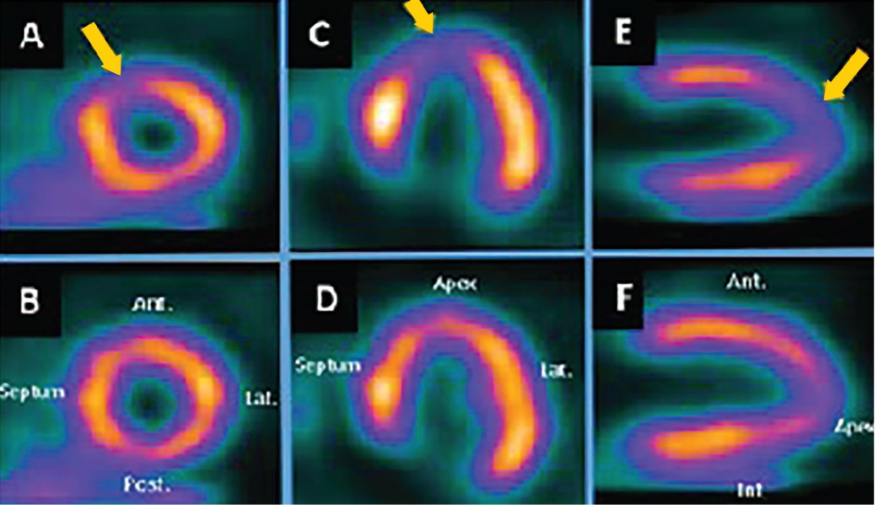
SPECT MPI showing a moderate-sized, reversible perfusion defect in the apex and septum (arrows in A, C, E), consistent with ischemia in the LAD territory. The rest images (B, D, F) show normal perfusion, confirming reversibility.
- Normal Scan: Homogeneous tracer uptake at both rest and stress.
- Reversible Defect (Ischemia): A region with reduced tracer uptake on the stress images that "fills in" and looks normal on the rest images. This indicates a lack of blood flow under stress that is restored at rest.
- Fixed Defect (Infarct/Scar): A region with reduced tracer uptake that is present on both the stress and rest images. This indicates non-viable heart muscle.
- Transient Ischemic Dilation (TID): The left ventricle appears larger on the stress images than at rest. This is a sign of severe, extensive ischemia.
3. Clinical Implications and Test Selection
The results of stress testing have profound implications for patient prognosis and management.
Prognostic Power:
- A normal MPI scan is associated with a very low risk (<1% per year) of major cardiac events (death or MI). This is often referred to as a "warranty period" of 1-2 years.
- The risk of future events increases directly with the extent and severity of ischemia found on the scan. A large, severe reversible defect identifies a high-risk patient who may benefit from revascularization.
Stress Nuclear: Methodology and Case Studies (Faisal Nabi, MD) January 28, 2020
Let's explore this prognostic power in more detail.
Watch the segment from 36:30 to 44:00. This is a crucial part of the lecture. Focus on: The excellent prognosis associated with a normal scan. How risk increases with the severity of perfusion abnormalities and worsening ejection fraction. The concept of the 'warranty period'.
Which Test Should You Choose?
The selection of the right test depends on the patient's clinical characteristics and the pre-test probability of disease.
-
Low-to-Intermediate Risk Patient, Able to Exercise, Interpretable ECG:
- EST is an excellent and cost-effective first choice.
-
Patient Unable to Exercise OR has an Uninterpretable Baseline ECG (e.g., LBBB, paced rhythm):
- Pharmacologic MPI is the test of choice. (Stress echocardiography is another valid option here).
-
Intermediate-to-High Risk Patient or Inconclusive Previous Test:
- MPI (exercise or pharmacologic) is often preferred for its higher diagnostic accuracy and powerful prognostic information.
-
Patient with Obesity:
- PET-MPI is superior to SPECT-MPI due to better image quality and less attenuation artifact, though its availability is limited.
Test your understanding!
A 68-year-old woman with a history of hypertension and diabetes presents with exertional dyspnea. Her baseline ECG shows a left bundle branch block (LBBB). She reports she can walk for about 5 minutes before getting short of breath. Which of the following is the most appropriate initial non-invasive test to evaluate for CAD?
- Standard Exercise Stress Test (EST)
- Dobutamine Stress Echocardiogram
- Regadenoson Myocardial Perfusion Imaging (MPI)
- Coronary CT Angiography
Show answer
The correct answer is 3. Regadenoson Myocardial Perfusion Imaging (MPI). The patient's baseline ECG has an LBBB, which makes a standard EST uninterpretable. Therefore, an imaging-based stress test is required. Between the imaging options, a vasodilator stress MPI (with Regadenoson or Adenosine) is generally preferred over dobutamine in patients with LBBB to avoid false-positive septal defects.
Conclusion
You have now learned how to interpret and apply two of the most important functional tests in cardiology. This knowledge allows you to move beyond anatomy and biomarkers to understand the physiological impact of coronary artery disease.
Key Takeaways:
- Exercise Stress Testing (EST) evaluates for ischemia based on ECG changes, functional capacity (METs), and hemodynamic response. Its main limitations are baseline ECG abnormalities and the patient's ability to exercise.
- Myocardial Perfusion Imaging (MPI) uses radiotracers to directly visualize blood flow. It is more sensitive than EST and can be performed with either exercise or pharmacologic stress.
- The key interpretation in MPI is differentiating a reversible defect (ischemia) from a fixed defect (infarct/scar).
- Both tests provide powerful prognostic information. A normal stress test (especially MPI) confers an excellent short-to-medium-term prognosis. The degree of abnormality directly correlates with future risk.
- Test selection is guided by the patient's ability to exercise and their baseline ECG.
In our last few lessons, we've built a robust diagnostic toolkit. You can interpret clinical findings, ECGs, chest X-rays, echos, biomarkers, and now, functional stress tests.
This lesson concludes the "Foundations in Clinical Cardiology and Diagnostics" module. You are now well-prepared to diagnose and risk-stratify patients. In our next lesson, we will begin the "Ischemic Heart Disease and Prevention" module. We will start at the very beginning by exploring the pathophysiology of atherosclerosis, the underlying disease process that these stress tests are designed to detect.