Hello! Welcome to the final lesson in our module, "Foundations in Clinical Cardiology and Diagnostics."
In our last session, we delved into non-invasive functional testing with exercise stress tests and myocardial perfusion imaging, learning how to detect the physiological consequences of coronary artery disease (CAD). Today, we move to the definitive anatomical test: invasive coronary angiography. This procedure is often considered the "gold standard" for visualizing the coronary arteries.
This lesson is designed to help you master the learning outcome: Describe the indications for and interpret findings of coronary angiography.
We will cover:
- The indications for coronary angiography: When is it appropriate to proceed with this invasive test, both in acute and chronic settings?
- The fundamentals of angiographic interpretation: You'll learn the standard views, coronary anatomy, and how to identify key structures.
- How to assess coronary lesions: We'll go beyond simple percentage stenosis to discuss flow dynamics (TIMI grade), physiological significance (FFR), and complex lesion characteristics.
- Integrating findings into clinical decisions: We'll touch upon scoring systems like the SYNTAX score that help guide the choice between treatments like PCI and CABG.
By the end of this lesson, you will have a comprehensive understanding of why and how coronary angiography is used, completing your foundational toolkit for diagnosing heart disease.
1. When is Coronary Angiography Indicated?
Deciding to proceed to invasive coronary angiography (ICA) is a critical step in a patient's management. The indications are based on a careful synthesis of the patient's symptoms, risk factors, and the results of non-invasive testing. We can broadly categorize them into acute and non-acute (chronic/stable) scenarios.
Acute and High-Risk Scenarios (Direct to Angiography)
In certain high-risk situations, we bypass further non-invasive testing and proceed directly to angiography.
Chest Pain Guideline Update: From AHA/ACC to ESC
The 2021 AHA/ACC chest pain guidelines clearly outline scenarios where direct invasive angiography is the recommended pathway. This short clip from a University of Washington Cardiology update summarizes these key points.
Watch the segment from 21:03 to 22:18. Focus on the four main criteria presented for considering direct invasive angiography.
As the video highlighted, direct ICA is indicated for:
- Acute Coronary Syndromes (ACS): Especially in STEMI (for primary PCI) and high-risk NSTEMI/UA (confirmed by troponins or high-risk scores like GRACE/TIMI).
- Newly Reduced Left Ventricular Ejection Fraction (LVEF): In patients with new, unexplained heart failure or reduced systolic function, as ischemia is a primary reversible cause.
- Hemodynamic Instability or Life-Threatening Arrhythmias: This includes cardiogenic shock, sustained ventricular tachycardia, or survivors of sudden cardiac death, where a critical coronary lesion is highly suspected.
- High-Risk Clinical Features: Based on validated risk scores or a constellation of findings suggesting a very high probability of severe, multivessel disease.
Stable/Chronic Scenarios (After Initial Evaluation)
In patients with stable symptoms, angiography is typically performed after non-invasive testing has identified high-risk features, or when symptoms persist despite optimal medical therapy.
Clinical Guideline for Diagnosis of Stable Ischemic Heart Disease
For a structured, recommendation-based view, let's review the guidelines for stable ischemic heart disease (IHD). This document provides very clear, testable points on when to use angiography.
Please read the recommendations under the main heading 'Coronary Angiography'. First, focus on 'Coronary Angiography as an Initial Testing Strategy' (Recommendations 21-23). Note how this overlaps with the high-risk scenarios we just discussed. Then, read 'Coronary Angiography to Assess Risk After Initial Workup' (Recommendations 24-28). Pay close attention to both the indications (Rec 24) and the important contraindications (Rec 25-28), which specify when angiography is not beneficial.
To summarize the key indications in stable patients:
- High-risk findings on non-invasive tests: Such as a large area of moderate-to-severe ischemia on MPI, significant abnormalities on stress echo, or high-risk coronary anatomy on CCTA (e.g., >50% left main stenosis).
- Lifestyle-limiting angina despite Guideline-Directed Medical Therapy (GDMT): When symptoms are not controlled with medications, angiography is performed to assess suitability for revascularization (PCI or CABG) to improve quality of life.
- Inconclusive non-invasive test results: When prior tests are ambiguous, angiography can provide a definitive diagnosis.
2. How to Read a Coronary Angiogram: Anatomy and Views
Interpreting a coronary angiogram is a visual skill built on a solid understanding of anatomy and radiographic projections. Let's build that foundation.
How to read a coronary angiography
This video, 'How to read a coronary angiography', is an excellent, in-depth tutorial. We will watch it in sections. This first part covers the absolute essentials: coronary anatomy, angiographic views, and how to orient yourself.
Watch the segment from 03:02 to 19:16. This is a detailed section, so focus on grasping these core concepts: The basic anatomy of the Left Main (LM), Left Anterior Descending (LAD), Left Circumflex (LCx), and Right Coronary Artery (RCA). The two main directions of movement for the C-arm: Right/Left Anterior Oblique (RAO/LAO) and Cranial/Caudal. How to use anatomical markers (diaphragm, catheter position, spine) to identify the view. The classic appearances of key views, such as the LAO Caudal ('Spider view') which is excellent for visualizing the left main bifurcation.
After watching that segment, you should be familiar with the main vessels and views. Here is a labeled image of a normal left coronary system in the LAO caudal view, often called the "spider view," which clearly shows the left main trifurcating.

3. Interpreting Pathological Findings
Now that you can identify the normal structures, let's learn how to describe the pathology.
Vessel Segmentation and Dominance
To report findings accurately, we divide each major artery into segments. We also determine the coronary "dominance," which refers to the artery that supplies the posterior descending artery (PDA) and posterolateral (PL) branches.
How to read a coronary angiography
Let's return to the tutorial video to learn about segmentation and dominance. These are fundamental concepts for describing lesion locations and understanding circulatory patterns.
Watch the segment from 23:45 to 29:34. Pay close attention to: How the LAD, LCx, and RCA are divided into proximal, mid, and distal segments. The definition of Right dominance (PDA from RCA, ~85% of people), Left dominance (PDA from LCx, ~8%), and Co-dominance.
Assessing Lesion Severity and Flow
Simply identifying a narrowing isn't enough. A full interpretation involves grading the stenosis, assessing its physiological impact, and evaluating blood flow.
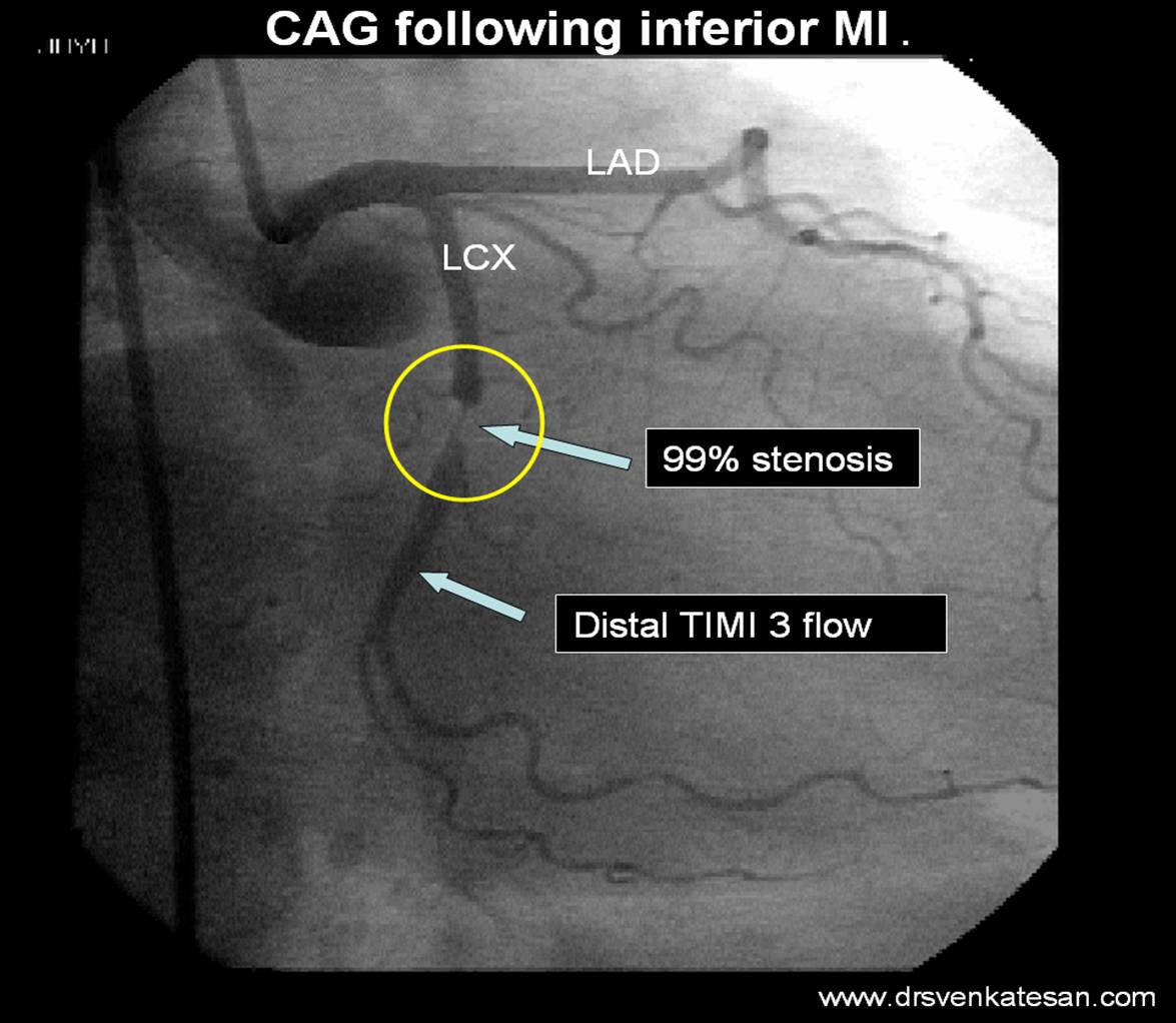
This image perfectly illustrates two key concepts: anatomical stenosis and functional flow.
How to read a coronary angiography
The next two sections of the video explain how to grade lesions and assess flow. This includes moving beyond simple visual estimation to more objective and physiological measures.
Watch from 29:14 to 39:49. This is a crucial section for your clinical and exam knowledge. Focus on: Lesion Grading: The difference between mild, moderate, and severe stenosis by visual estimation. Fractional Flow Reserve (FFR): The principle of this physiological measurement and the critical cutoff value of ≤ 0.80 indicating a hemodynamically significant stenosis. TIMI Flow Grade: Understand the scale from 0 (no flow) to 3 (normal flow). This is essential for assessing perfusion, especially in ACS.
A key takeaway from the video is the finding from the FAME trial: an angiographically intermediate lesion (50-70%) is only hemodynamically significant by FFR about 35% of the time. This highlights the limitation of relying on anatomy alone and the importance of physiological assessment.
Test your understanding!
A patient with stable angina undergoes angiography. You find a 60% stenosis in the mid-LAD. The cardiologist performs an FFR measurement across the lesion, which yields a value of 0.88. What is the clinical significance of this finding?
Show answer
The FFR value of 0.88 is greater than the ischemic threshold of 0.80. This means that despite the moderate anatomical appearance of the stenosis, it is not causing a significant limitation to blood flow (i.e., it is not hemodynamically significant). Therefore, per current guidelines, this lesion should be treated medically, and stenting (PCI) would not be indicated. This demonstrates the critical role of physiological testing in guiding revascularization decisions.
Specific Lesion Types and the SYNTAX Score
Finally, a complete interpretation involves characterizing the type of lesion and, in complex cases, using a scoring system to guide management.
How to read a coronary angiography
To round out your interpretation skills, let's cover some special lesion types and a critical decision-making tool, the SYNTAX score.
Watch the final recommended segment from 39:31 to 50:23. Focus on: Recognizing the angiographic appearance of thrombus, calcification, and myocardial bridges. Understanding the purpose of the SYNTAX score: to quantify the complexity of coronary disease and help the Heart Team decide between PCI and CABG, especially in patients with left main or multivessel disease.
The SYNTAX score is a powerful tool. A high score (e.g., >33 in left main disease, or >22 in three-vessel disease) suggests that CABG offers a better long-term outcome than PCI. This integration of anatomical findings into a risk score is a cornerstone of modern interventional cardiology.
Conclusion
Congratulations on completing the "Foundations in Clinical Cardiology and Diagnostics" module! You have systematically built a comprehensive diagnostic skillset, from clinical examination and ECGs to advanced functional and anatomical imaging.
Key Takeaways from this Lesson:
- Coronary Angiography (ICA) is the gold standard for defining coronary anatomy, with clear indications for its use in both acute and stable settings.
- Interpretation requires a systematic approach: identifying views, understanding coronary anatomy and dominance, and describing lesions by location (segment) and severity (% stenosis).
- Physiological Assessment with FFR is crucial for determining the significance of intermediate lesions and guiding revascularization.
- Flow Assessment using the TIMI grade is vital for evaluating myocardial perfusion, particularly in ACS.
- Clinical Decision-Making is guided by integrating anatomical findings (e.g., using the SYNTAX score) to determine the optimal treatment strategy (medical therapy, PCI, or CABG).
In our next lesson, we will begin a new module: "Ischemic Heart Disease and Prevention." Having mastered the tools to diagnose CAD, we will now go back to the very beginning and explore the underlying disease process itself, starting with the pathophysiology of atherosclerosis.