Hello! Welcome to your next lesson in the "Foundations in Clinical Cardiology and Diagnostics" module.
In our last lesson, we focused on clinical synthesis, applying diagnostic algorithms for the common presentations of chest pain, dyspnea, and palpitations. You learned that a crucial step in these algorithms is often ordering cardiac biomarkers to clarify the diagnosis. Today, we will dive deep into those biomarkers.
This lesson is designed to help you master the learning outcome: Integrate cardiac biomarkers (Troponins, BNP/NT-proBNP) into the diagnostic and prognostic evaluation of cardiac patients.
We will explore the two most important groups of cardiac biomarkers in modern cardiology:
- Cardiac Troponins (cTn): The cornerstone for diagnosing myocardial injury and infarction.
- Natriuretic Peptides (BNP and NT-proBNP): Essential for the diagnosis and management of heart failure.
For each, we'll cover their underlying pathophysiology, their kinetics, and most importantly, how to interpret their levels within the full clinical context to make accurate diagnostic and prognostic assessments. This knowledge is not only vital for clinical practice but is also a high-yield topic for your NEET-PG and university examinations.
1. Cardiac Troponins: Markers of Myocardial Injury
When you suspect Acute Coronary Syndrome (ACS) in a patient with chest pain, cardiac troponins are the go-to blood test. Let's understand why.
Pathophysiology and Kinetics
Cardiac troponins (cTn) are proteins that are part of the contractile apparatus of heart muscle cells. Specifically, we measure two isoforms that are unique to the heart: cardiac troponin I (cTnI) and cardiac troponin T (cTnT).
When myocardial cells are irreversibly injured (necrosis), their membranes rupture, releasing these proteins into the bloodstream where they can be detected.
Cardiovascular Biomarkers: Tools for Precision Diagnosis
To start, let's review the fundamental properties of troponins and their role as the gold-standard biomarker for diagnosing acute myocardial infarction (AMI).
Read the section titled 'Cardiac troponin I (cTnI) and cardiac troponin T (cTnT)'. Focus on why they are considered the gold standard and the basic principle of their release during myocardial necrosis.
A critical concept in diagnosing an acute event like an MI is the dynamic change in troponin levels. A single elevated value is not enough. In a classic MI, troponin levels follow a predictable pattern:
- They begin to rise 2-4 hours after the onset of injury.
- They peak at around 12-24 hours.
- They gradually return to baseline over several days (up to 7-14 days for cTnT).
This characteristic "rise and/or fall" is essential for differentiating an acute event from chronic myocardial injury.
The Era of High-Sensitivity Troponins (hs-cTn)
Modern medicine uses high-sensitivity cardiac troponin (hs-cTn) assays. These assays are a significant advancement because they can detect much lower concentrations of troponin and with greater precision than older tests. This has several important implications for clinical practice.
Learn to Love High Sensitivity Troponins | The EM & Acute Care Course
This video, titled 'Learn to Love High Sensitivity Troponins', explains the practical impact of these advanced assays. It discusses how they allow for faster decision-making in the emergency department.
Watch the following segments: 00:00 - 03:06: Understand the difference in sensitivity compared to regular troponins and how hs-cTn can help rule out significant coronary events rapidly. 03:06 - 05:48: Focus on the importance of the 'delta' (change in levels) and the concept of ruling out an MI with a single test in very low-risk patients. 10:37 - 14:10: Pay attention to the different rule-out protocols (e.g., 0 and 1-hour, 0 and 3-hour) and how they are applied. Note that the speaker's style is quite dynamic and opinionated, but the core clinical points about rapid protocols are extremely valuable for your exams.
Key takeaways from hs-cTn:
- Higher Sensitivity: Can detect troponin in most healthy individuals, establishing a normal range. The diagnostic threshold is set at the 99th percentile of a healthy population.
- Faster Protocols: The precision of hs-cTn allows for the use of rapid diagnostic algorithms (e.g., 0/1-hour or 0/3-hour protocols), where a second sample is drawn much sooner. This significantly speeds up the rule-in or rule-out of MI in the emergency department.
- Negative Predictive Value: A very low or "undetectable" hs-cTn level on presentation has an excellent negative predictive value, meaning it can confidently rule out MI in many patients, allowing for safe and early discharge.
Integrating Troponins: Beyond Myocardial Infarction
An elevated troponin indicates myocardial injury. It does not automatically mean the patient is having a classic heart attack (Type 1 Myocardial Infarction). The art of clinical medicine is to interpret the elevated troponin in the context of the patient's history, exam, and ECG findings.
The following flowchart is the key to structured thinking when you get an elevated troponin report.

Let's break down the conditions in this algorithm:
- Acute Myocardial Infarction (AMI): Defined as acute myocardial injury (a rise and/or fall of troponin) in the setting of clinical evidence of acute myocardial ischemia (e.g., ischemic symptoms, new ECG changes, new wall motion abnormality on echo).
- Type 1 MI: The classic heart attack caused by atherosclerotic plaque rupture and coronary thrombosis.
- Type 2 MI: An MI due to an oxygen supply-demand mismatch (e.g., severe anemia, tachyarrhythmia, shock) in the absence of acute plaque rupture. The underlying coronary arteries might have stable plaque, but the acute stress is what tips the balance into ischemia and necrosis.
- Acute Myocardial Injury: A rise and/or fall of troponin, but without clinical evidence of ischemia. This is seen in conditions like myocarditis or after a procedure like cardioversion.
- Chronic Myocardial Injury: Persistently elevated, stable troponin levels without a significant rise or fall. This is common in patients with structural heart disease or chronic kidney disease (CKD).
The following video provides excellent case-based examples that illustrate these distinctions.
Elevated Troponins: Is It an MI or Not? Key Insights and Case Examples
The video 'Elevated Troponins: Is It an MI or Not?' walks through several clinical cases to demonstrate how to differentiate between MI and non-MI causes of troponin elevation. This is a perfect way to apply the flowchart we just saw.
Watch the case examples from 02:33 to 06:22 and from 08:09 to 12:28, and finally 15:33 to 16:45. For each case (myocarditis, aortic dissection, heart failure, renal failure/hyperkalemia, stroke, pancreatitis, PE), note the following: The clinical presentation (symptoms, exam, ECG). The troponin pattern (significant rise/fall vs. stable/modest change). The final diagnosis and why it explains the troponin elevation.
Prognostic Value of Troponins:
An important point is that any detectable myocardial injury, even if chronic, is prognostically significant. Patients with CKD or stable heart failure who have chronically elevated troponins have a higher risk of future adverse cardiovascular events and mortality compared to similar patients with normal troponins.
Test your understanding!
A 70-year-old man with end-stage renal disease (ESRD) on hemodialysis is admitted for a non-healing foot ulcer. A routine blood test shows a troponin I level that is three times the upper limit of normal. His previous troponin level from three months ago was similar. The patient has no chest pain, and his ECG is unchanged from baseline. According to the diagnostic algorithm, how would you classify this troponin elevation?
Show answer
This is classified as Chronic Myocardial Injury. The troponin level is elevated but stable (no significant rise or fall), and there is no clinical evidence of acute myocardial ischemia. This pattern is common in patients with ESRD due to a combination of reduced clearance and underlying uremic cardiomyopathy. While not an acute MI, it does indicate a higher long-term cardiovascular risk.
2. Natriuretic Peptides: Markers of Myocardial Stretch
When a patient presents with dyspnea, a key question is whether the cause is cardiac or pulmonary. Natriuretic peptides are invaluable in making this distinction.
Pathophysiology and Biochemistry
The two main natriuretic peptides used in clinical practice are B-type natriuretic peptide (BNP) and N-terminal pro-B-type natriuretic peptide (NT-proBNP).
Biomarkers in acute heart failure
To understand how to use these markers, we first need to know where they come from and what they do. This text from an Oxford textbook on acute heart failure provides a clear explanation.
Read the sections 'Natriuretic peptides' and 'Natriuretic peptide biochemistry'. Focus on: The stimulus for release (ventricular stretch). The precursor molecule (proBNP) and how it's cleaved into active BNP and inactive NT-proBNP. The key differences in their half-life and clearance.
In essence, when the ventricles are stretched due to high pressure or volume overload (the cardinal features of heart failure), the heart releases proBNP, which is cleaved into BNP and NT-proBNP. BNP is a biologically active hormone that promotes vasodilation and natriuresis (excretion of sodium and water) to counteract the volume overload. NT-proBNP is an inactive fragment.
Because NT-proBNP has a longer half-life and is more stable in a blood sample, it is often preferred in clinical practice.
Diagnostic and Prognostic Use in Heart Failure
The primary use of natriuretic peptides is in the evaluation of a patient with acute dyspnea.
- Diagnosis: A very low BNP or NT-proBNP level makes a diagnosis of acute heart failure highly unlikely. Conversely, a very high level strongly supports it.
- Prognosis: The level of natriuretic peptide correlates with the severity of heart failure and is a powerful predictor of mortality and rehospitalization.
- Therapy Guidance: A significant drop in natriuretic peptide levels during treatment for acute decompensated heart failure indicates a good response to therapy (e.g., diuresis).
Interpreting Natriuretic Peptide Levels
Like troponins, natriuretic peptide levels must be interpreted with care, considering the full clinical picture.
Biomarkers in acute heart failure
This next section from the same resource discusses the practical challenges of interpretation, including 'grey zones' and other conditions that can affect the results.
Read the section 'Interpreting natriuretic peptide levels' and review the associated tables (Table 37.2 and 37.3). Pay close attention to: The concept of 'grey zone' values, where the diagnosis is uncertain. Factors that can falsely increase NP levels (e.g., renal failure, atrial fibrillation, old age). The one major factor that can falsely lower NP levels (obesity). The important effect of sacubitril/valsartan (Entresto) on BNP vs NT-proBNP.
This last point is crucial for your exams:
- Sacubitril is a neprilysin inhibitor. Neprilysin is an enzyme that breaks down BNP.
- Therefore, in a patient taking sacubitril/valsartan, BNP levels will be artificially high and cannot be used to monitor HF status.
- NT-proBNP is not broken down by neprilysin, so its levels will accurately reflect the patient's hemodynamic stress. You must use NT-proBNP to monitor these patients.
Test your understanding!
An 80-year-old woman with a history of atrial fibrillation and moderate chronic kidney disease presents with acute dyspnea. Her NT-proBNP level is elevated but falls into the "grey zone" for her age. What are three factors in her history that could be contributing to her NT-proBNP level, making the diagnosis of acute heart failure less certain based on this value alone?
Show answer
The three factors are:
- Advanced Age: NT-proBNP levels naturally increase with age.
- Atrial Fibrillation: AF causes atrial and ventricular stretch, leading to elevated natriuretic peptide levels even without overt heart failure.
- Chronic Kidney Disease: NT-proBNP is cleared by the kidneys, so impaired renal function leads to higher baseline levels.
3. The Integrated Biomarker Landscape
While troponins and natriuretic peptides are the workhorses, they are part of a larger family of biomarkers that reflect different aspects of cardiac pathology.
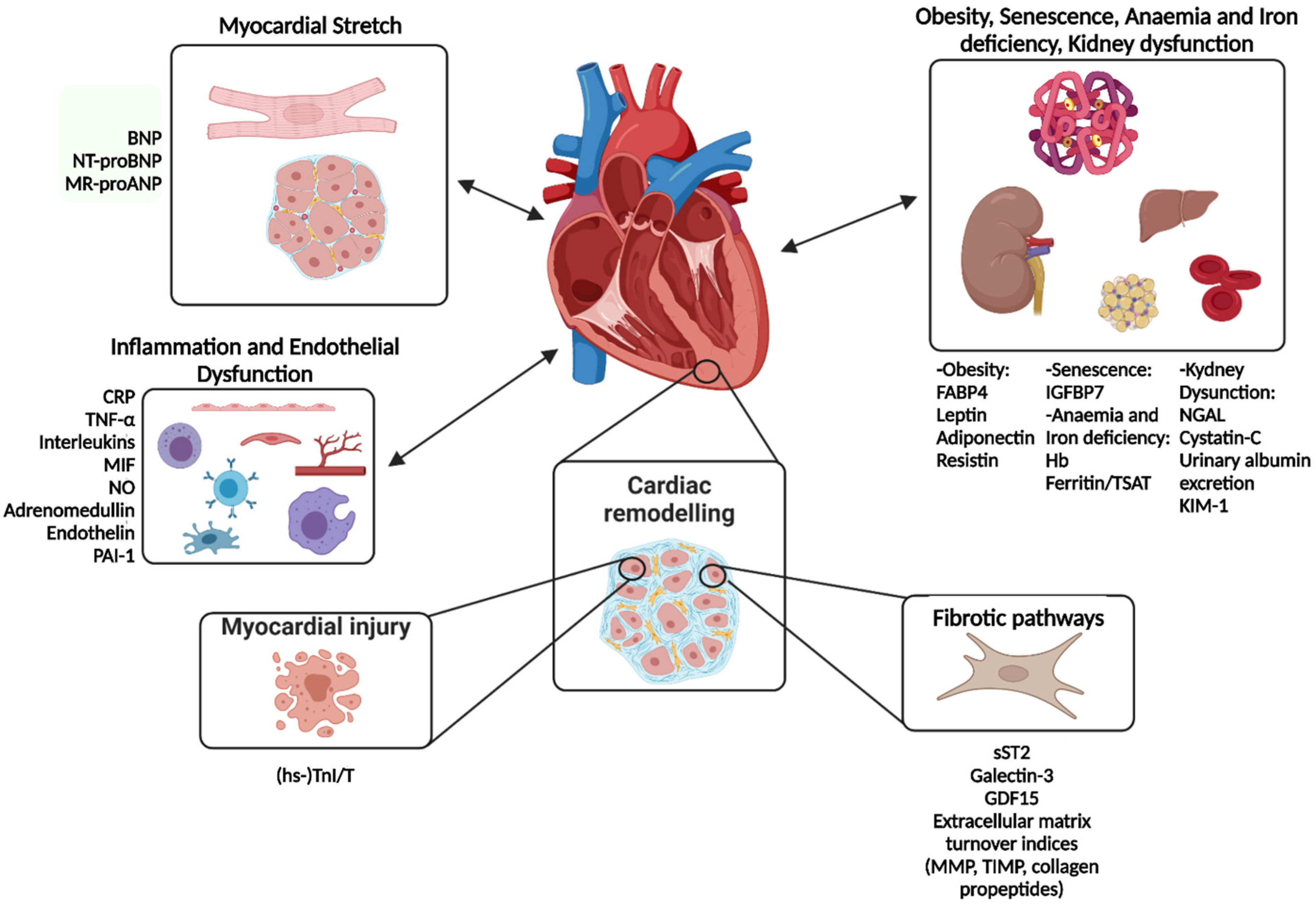
This image reinforces our main points: troponins reflect injury, and natriuretic peptides reflect stretch. While a deep dive into other markers like ST2 (fibrosis/remodeling) or hs-CRP (inflammation) is beyond today's core objective, it's important to recognize that a multi-marker approach is the future of cardiovascular diagnostics. For your reference, Table 4 in the Cardiovascular Biomarkers: Tools for Precision Diagnosis article provides a comprehensive summary of many of these markers.
Conclusion
You have now completed a thorough review of the two most important biomarkers in clinical cardiology. Integrating these powerful tools correctly into your diagnostic reasoning is a skill that separates the novice from the expert.
Key Takeaways:
- Troponin signifies myocardial injury. The diagnosis of Myocardial Infarction requires a dynamic change (rise and/or fall) in the context of clinical evidence of ischemia.
- Always consider the broad differential for an elevated troponin, including Type 2 MI and non-ischemic myocardial injury (e.g., myocarditis, PE, sepsis, CKD).
- High-sensitivity troponins enable rapid rule-in/rule-out protocols, transforming emergency care for chest pain.
- BNP/NT-proBNP signify myocardial stretch. They are excellent for diagnosing heart failure in patients with dyspnea and for determining prognosis.
- Always interpret biomarker values in the context of the patient's age, renal function, body weight, and comorbidities.
In this module, we have built your foundational diagnostic toolkit. You can now take a history, perform an exam, and interpret an ECG, CXR, Echo, and key biomarkers. You are ready to start diagnosing specific cardiac diseases.
Our next lesson will be the first in a new module, "Ischemic Heart Disease and Prevention." We will begin by exploring the fundamental process that leads to most myocardial infarctions: the pathophysiology of atherosclerosis. Understanding how stable plaques form and progress to unstable, thrombotic lesions will connect perfectly with what you've learned today about detecting the downstream consequence—myocardial injury.