Hello! Welcome to your fifth lesson in the Foundations in Clinical Cardiology and Diagnostics module.
In our previous lessons, we've systematically built your diagnostic toolkit. You've learned to interpret findings from the clinical examination, the 12-lead ECG, chest X-rays, and most recently, the detailed structural and functional data from an echocardiogram. Now, it's time to put it all together.
This lesson serves as a capstone for the module, focusing on clinical synthesis. A patient doesn't present with "Grade 2 diastolic dysfunction"; they present with shortness of breath. Our goal today is to bridge the gap between patient symptoms and diagnostic findings. We will directly address the learning outcome: to apply diagnostic algorithms for the evaluation of common cardiac presentations: chest pain, dyspnea, and palpitations.
For each of these three cardinal symptoms, we will work through a systematic, evidence-based algorithm that integrates history, physical exam, and the diagnostic tests you've studied. This approach is fundamental for your clinical practice and crucial for your exam preparation, mirroring the structured thinking required to solve complex clinical vignettes.
1. The Diagnostic Algorithm for Chest Pain
Chest pain is one of the most common and high-stakes presentations in medicine. The primary goal is to quickly identify and manage life-threatening conditions. A structured approach is not just helpful; it's essential.
Initial Triage and the "Big Six"
The first step, always, is to assess the patient's stability using the ABCDE (Airway, Breathing, Circulation, Disability, Exposure) approach. An unstable patient requires immediate stabilization, often concurrently with the diagnostic workup.
The video below provides an excellent overview of the systematic approach, focusing on rapidly ruling out the most dangerous causes of chest pain.
Approach to chest pain: Clinical sciences
This video from Osmosis, 'Approach to chest pain', outlines a clear, step-by-step method for evaluating a patient with chest pain, emphasizing the immediate need to identify life-threatening conditions.
Please watch the video from the beginning up to 11:08. As you watch, create a mental checklist of the key features (history, exam, ECG, imaging) for each of these six life-threatening conditions: Acute Coronary Syndrome (STEMI) Cardiac Tamponade Aortic Dissection Pulmonary Embolism (PE) Tension Pneumothorax Esophageal Rupture
The Diagnostic Pathway in Detail
Let's formalize the algorithm discussed in the video, incorporating key guidelines and the tools we've learned.
Chest Pain Evaluation: Updated Guidelines From the AHA...
This article from American Family Physician summarizes the 2021 AHA/ACC guidelines for chest pain evaluation. It reinforces the video's concepts and adds important details on modern diagnostic strategies.
Read the sections 'Key Points for Practice,' 'Initial Evaluation,' and 'Diagnostic Evaluation.' Pay close attention to: The recommendation to obtain a 12-lead ECG within 10 minutes. The move away from the term 'atypical' chest pain. The role of high-sensitivity cardiac troponin (hs-cTn).
Here is a summary of the diagnostic algorithm for acute chest pain:
-
Assess Stability (ABCDE): Stabilize if needed. Provide oxygen if hypoxemic, establish IV access, and place the patient on a cardiac monitor.
-
Initial Evaluation (within 10 minutes):
- Focused History: Use OPQRST/SOCRATES to characterize the pain. Remember that classic "cardiac" features (pressure, squeezing, radiating) increase suspicion, but their absence, especially in women, the elderly, and diabetics, does not rule out ACS.
- Focused Physical Exam: Look for signs of hemodynamic instability (hypotension, tachycardia), heart failure (crackles, elevated JVP), or specific clues like asymmetric pulses (aortic dissection) or a new murmur.
- 12-Lead ECG: This is the most critical initial test. The primary goal is to identify ST-elevation myocardial infarction (STEMI) or a new Left Bundle Branch Block (LBBB), which require immediate reperfusion therapy.
-
Interpreting Initial Results & Next Steps:
- If ECG shows STEMI: The diagnosis is made. The patient needs to go to the cardiac cath lab for primary PCI or receive thrombolysis.
- If ECG is non-diagnostic for STEMI: The workup continues.
- Measure Cardiac Troponins: Use a high-sensitivity troponin assay. A second level is checked 1-3 hours later. Elevated troponins in the right clinical context diagnose a Non-ST-Elevation Myocardial Infarction (NSTEMI). Normal troponins but high clinical suspicion may indicate Unstable Angina (UA).
- Consider Other Life-Threatening Causes:
- Aortic Dissection: Suspect with tearing pain radiating to the back, pulse deficits, or a widened mediastinum on CXR. Confirm with CT Angiogram (if stable) or a transesophageal echo (if unstable).
- Pulmonary Embolism: Suspect with pleuritic chest pain, dyspnea, and hypoxia. Use a risk score (like the Wells score) to guide testing with D-dimer or CT Pulmonary Angiogram.
- Cardiac Tamponade: Suspect with Beck's triad (hypotension, JVD, muffled heart sounds). ECG may show low voltage and electrical alternans. Confirm with an urgent echocardiogram.
- Tension Pneumothorax: A clinical diagnosis based on respiratory distress, unilateral absent breath sounds, tracheal deviation, and hypotension. Do not wait for a CXR; perform immediate needle decompression.
-
Risk Stratification for Possible ACS:
- For patients where ACS is suspected but not confirmed, we use clinical decision pathways like the HEART score (History, ECG, Age, Risk factors, Troponin).
- Low-risk patients (e.g., HEART score 0-3) can often be safely discharged from the emergency department without further testing.
- Intermediate-risk patients may undergo non-invasive testing like cardiac CT angiography (CCTA) or stress testing to look for underlying coronary artery disease (CAD).
Test your understanding!
A 68-year-old man with a history of poorly controlled hypertension presents to the emergency department with a sudden onset of severe, "tearing" chest pain that radiates to his back between the shoulder blades. His BP is 190/110 mmHg in the right arm and 140/90 mmHg in the left. His ECG shows sinus tachycardia with LVH but no acute ST changes. A chest X-ray reveals a widened mediastinum. What is the most likely diagnosis, and what is the single most appropriate next diagnostic test if the patient is hemodynamically stable?
Show answer
The most likely diagnosis is Aortic Dissection. The classic "tearing" pain radiating to the back, history of hypertension, significant blood pressure differential between arms, and widened mediastinum on CXR are all highly suggestive.
The most appropriate next diagnostic test in a stable patient is a CT Angiogram of the chest and aorta.
2. The Diagnostic Algorithm for Dyspnea
Dyspnea, or shortness of breath, is another common symptom with a wide differential spanning cardiac, pulmonary, and systemic causes. Like chest pain, the initial approach must be swift and systematic.
A Pathophysiological Framework
Before jumping into a list of diseases, it's helpful to think about the underlying physiological reasons a person feels short of breath.
This video, 'An Approach to Acute Dyspnea' from Strong Medicine, provides an excellent framework for diagnosis, starting from pathophysiology and moving to a practical, step-by-step clinical algorithm.
Please watch from the beginning to 3:24 to understand the pathophysiological triggers of dyspnea. Then, skip to 5:09 and watch until 12:20 to follow the practical diagnostic algorithm.
A Step-by-Step Diagnostic Pathway
The algorithm for dyspnea hinges on combining a few key pieces of data to narrow the possibilities. This flowchart, based on principles from Harrison's Manual of Medicine, visualizes this process.

Let's break down this pathway:
-
Initial Data Set:
- History: Onset (sudden vs. gradual), triggers (exertion, rest, position - orthopnea/PND), associated symptoms (chest pain, cough, fever, wheeze).
- Vital Signs: Pay close attention to respiratory rate, oxygen saturation, heart rate, and blood pressure.
- Physical Exam:
- Lungs: Listen for crackles (edema, pneumonia, fibrosis), wheezes (asthma, COPD), or absent breath sounds (pneumothorax, large effusion).
- Heart: Check for elevated JVP, an S3 gallop, new murmurs, or peripheral edema (all suggestive of heart failure).
- Chest X-Ray: This is a pivotal test. Look for cardiomegaly, pulmonary edema, focal consolidation, pleural effusion, or hyperinflation.
-
Branching Based on Findings: The combination of lung exam and CXR findings points toward specific diagnoses, as outlined in the Strong Medicine video:
- Focal crackles + Focal opacity on CXR -> Pneumonia.
- Diffuse crackles + Diffuse opacities on CXR -> Pulmonary Edema.
- Is it cardiogenic? Look for cardiac clues: elevated JVP, S3, edema, cardiomegaly on CXR, and a high BNP/NT-proBNP. An echocardiogram showing low LVEF or severe diastolic dysfunction (from our last lesson) confirms a cardiac origin.
- Decreased breath sounds + Pleural opacity on CXR -> Pleural Effusion.
- Wheezing + Normal/Hyperinflated CXR -> Asthma/COPD exacerbation.
-
The "Normal Exam, Normal CXR" Challenge: This is where the algorithm gets more interesting.
- If the patient is HYPOXEMIC: Your top suspicion must be Pulmonary Embolism. The absence of other findings to explain hypoxia is a major red flag for PE. Proceed with risk stratification (Wells score) and CTPA/D-dimer.
- If the patient is NOT hypoxemic: Cast a wider net.
- Myocardial Ischemia (Angina Equivalent): Dyspnea can be the sole presentation of ACS, especially in the elderly. Check ECG and troponins.
- Anemia: Check a complete blood count (CBC).
- Metabolic Acidosis: Check electrolytes and a blood gas. The patient may be breathing deeply (Kussmaul respirations) to compensate for acidosis (e.g., DKA).
- Anxiety/Panic Attack: This is a diagnosis of exclusion. Never assume anxiety without ruling out life-threatening organic causes.
Test your understanding!
A 72-year-old female presents with two days of progressive shortness of breath and a dry cough. Her vitals are: HR 110, BP 160/95, RR 28, SpO2 91% on room air. On exam, you note an elevated JVP to the angle of the jaw, bilateral crackles halfway up the lung fields, and 2+ pitting edema in her lower extremities. Her ECG shows sinus tachycardia. An echo from 6 months ago showed an LVEF of 35%. What is the most likely diagnosis?
Show answer
The most likely diagnosis is Acute Decompensated Heart Failure, presenting as acute cardiogenic pulmonary edema. The combination of risk factors (known HFrEF), physical exam findings (elevated JVP, lung crackles, peripheral edema), and vital signs (tachycardia, tachypnea, hypoxia) is classic for this presentation.
3. The Diagnostic Algorithm for Palpitations
Palpitations—an unpleasant awareness of one's own heartbeat—are very common. The challenge is to distinguish benign, isolated ectopics from potentially life-threatening arrhythmias. The history is paramount.
Initial Evaluation and Differential Diagnosis
The first step is to characterize the sensation and search for "red flags."
Outpatient Approach to Palpitations
This comprehensive article, 'Outpatient Approach to Palpitations', provides an excellent framework for evaluation, covering the differential diagnosis, key historical features, and the diagnostic testing pathway.
Read the article up to the 'Specific Cardiac Arrhythmias' section (pages 63-66). Focus on: The broad categories of causes: cardiac (structural/arrhythmic), psychiatric, and miscellaneous (Tables 1 & 3). Key historical clues and red flags (e.g., association with syncope). The role of the initial 12-lead ECG and when to proceed to ambulatory monitoring.
Key Historical Questions:
- How would you describe it? (fluttering, pounding, skipped beats) -> Have the patient tap it out.
- Is it fast or slow? Regular or irregular?
- When does it occur? (at rest, with exercise, after caffeine)
- Are there associated symptoms? Syncope (fainting) or near-syncope is a major red flag. Chest pain and severe dyspnea are also concerning.
- What is your personal and family history? (known heart disease, family history of sudden cardiac death).
The Diagnostic Pathway
The following algorithm provides a clear, step-by-step approach to investigation.
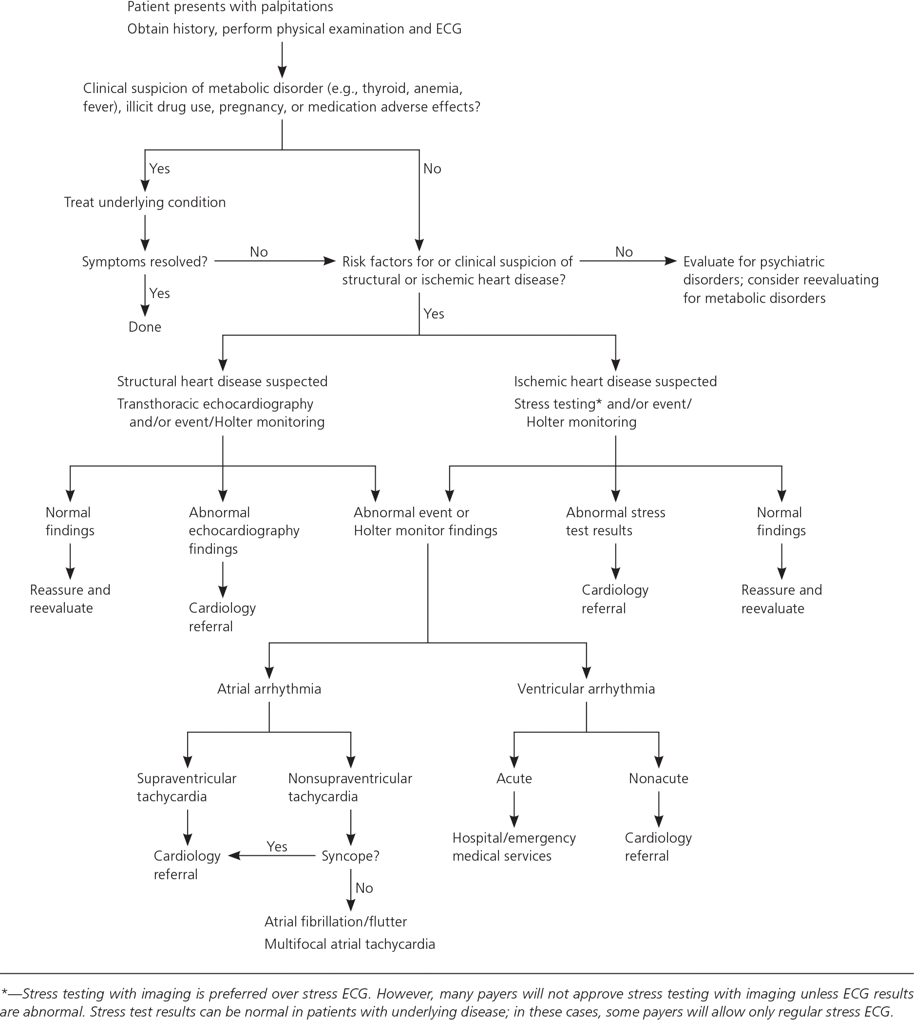
Let's walk through this algorithm:
-
Initial Evaluation:
- History & Physical Exam: As detailed above.
- 12-Lead ECG: Even a normal resting ECG is valuable. It can reveal underlying structural disease (e.g., LVH, Q waves from a prior MI) or an arrhythmic substrate (e.g., long QT interval, delta wave of Wolff-Parkinson-White syndrome).
- Basic Labs: Consider checking a TSH (for thyroid disease) and CBC (for anemia) based on clinical suspicion.
-
Interpreting Initial Findings:
- If a clear cause is found (e.g., hyperthyroidism, severe anemia, obvious arrhythmia on the initial ECG like atrial fibrillation), the diagnosis is made, and management can begin.
- If the initial evaluation is non-diagnostic (the most common scenario), the next steps depend on the frequency of symptoms and the presence of risk factors.
-
Further Investigation for Suspected Arrhythmia:
- Frequent Symptoms (daily): A 24-48 hour Holter monitor is appropriate. This continuously records the ECG, allowing correlation of symptoms with the underlying rhythm.
- Infrequent Symptoms (weekly/monthly): An event monitor or loop recorder is better. The patient can wear this for weeks and activate it to record the ECG when symptoms occur. Newer models can also auto-detect arrhythmias.
- Exertional Symptoms: An exercise stress test is the test of choice to try and provoke the arrhythmia under controlled conditions.
- Suspected Structural Disease: An echocardiogram is indicated if the exam suggests a murmur or if there are risk factors for structural heart disease.
-
Referral and Management:
- High-Risk Features: Patients with palpitations associated with syncope, a family history of sudden death, or known severe structural heart disease (e.g., severe LV dysfunction, hypertrophic cardiomyopathy) require urgent referral to a cardiologist, regardless of whether an arrhythmia has been captured.
- Benign Findings: If monitoring reveals isolated premature atrial or ventricular contractions (PACs/PVCs) in an otherwise healthy heart, the patient can be reassured and counseled on trigger avoidance (caffeine, alcohol).
- Specific Arrhythmia Diagnosed: If a sustained arrhythmia like SVT, atrial fibrillation, or ventricular tachycardia is captured, specific management and cardiology referral are required.
Test your understanding!
A 25-year-old medical student presents with palpitations that she describes as "fluttering" in her chest. They occur a few times a week, last for a few minutes, and are sometimes associated with a feeling of lightheadedness, but never true loss of consciousness. She has no significant past medical history and her family history is unremarkable. Her physical exam and initial 12-lead ECG are completely normal. What is the most appropriate next step in her management?
Show answer
The most appropriate next step is to order an ambulatory ECG monitor. Given that her symptoms are infrequent (a few times a week), a patient-activated event monitor or a loop recorder worn for 30 days would be more likely to capture an episode than a 24-hour Holter monitor. This will help determine if her symptoms correlate with a cardiac arrhythmia. Reassurance alone is not sufficient given the associated lightheadedness.
Conclusion
Congratulations on completing this crucial lesson. You have learned to apply structured, logical thinking to three of the most important presentations in cardiology. By starting with a systematic assessment and using diagnostic tools to answer specific questions, you can navigate complex clinical scenarios safely and efficiently.
Key Takeaways:
- Systematic Approach is Key: For chest pain, dyspnea, and palpitations, always begin with an assessment of stability (ABCs) and a thorough history and physical exam.
- Prioritize the Dangerous: Your initial workup should focus on ruling out immediate life-threatening conditions (e.g., STEMI, PE, Aortic Dissection).
- Integrate Your Tools: The ECG, CXR, echocardiogram, and biomarkers are not isolated tests. They are pieces of a puzzle that you must assemble to see the full clinical picture. The echo finding of an RV strain pattern supports a diagnosis of PE, while high E/e' explains the pulmonary edema seen on CXR.
- Algorithmic Thinking: Following established diagnostic pathways ensures a thorough workup, minimizes errors, and helps you decide on the next logical step, whether it's an urgent intervention, further testing, or safe discharge.
You have now completed the "Foundations" module. You are well-equipped with the fundamental diagnostic skills of a cardiologist. In our next module, we will begin our deep dive into specific disease states, starting with the pathophysiology of the most common culprit behind cardiac chest pain: Ischemic Heart Disease. Your work in this lesson has prepared you perfectly to understand how we diagnose this condition in the real world.