Welcome to your next lesson. In our last session, we established the fundamental kinetic principles of levers, torque, and force couples that govern human movement. Today, we will apply those concepts to one of the most intricate and vital structures in the body: the spinal column.
This lesson focuses on the biomechanics of the spine, specifically the function of the intervertebral discs and ligaments. A solid grasp of this topic is essential for your MPT entrance exams, as it forms the basis for assessing and managing a wide array of spinal conditions. We will explore how the spine masterfully balances the conflicting demands of providing rigid protection for the spinal cord while allowing for a remarkable degree of mobility.
1. The Functional Unit of the Spine: The Motion Segment
To understand the biomechanics of the spine, we first break it down into its smallest functional unit: the motion segment. This concept is central to analyzing spinal movement and pathology.
Biomechanics Lecture 4 - Spine
The "Rehab Science" channel provides an excellent introduction to the functional units and overall structure of the spine.
Please watch the segment from this video, which defines the motion segment and explains the development and purpose of the primary (kyphotic) and secondary (lordotic) spinal curves.
As the video explained, a motion segment consists of two adjacent vertebrae and the soft tissues connecting them, primarily the intervertebral disc (IVD) and the facet joints. The normal S-shaped curves of the spine (cervical and lumbar lordosis, thoracic kyphosis) are crucial for shock absorption, distributing axial loads more effectively than a straight, rigid rod.
The Intervertebral Disc (IVD)
The IVD is a remarkable structure, accounting for about 25% of the total height of the spinal column. It has two main components:
- Nucleus Pulposus: A gel-like central mass with high water content. It functions like a hydraulic ball-bearing, resisting compressive forces and allowing the vertebrae to pivot and rock on one another.
- Annulus Fibrosus: A series of tough, concentric fibrocartilage rings that encase the nucleus. The fibers in each layer are oriented obliquely, crisscrossing each other to provide formidable resistance to tensile (stretching) and torsional (twisting) forces.
Together, the IVDs serve four primary functions: absorbing shock, allowing mobility, maintaining the space between vertebrae for nerve root exit, and guiding motion.
2. The Passive Stabilizers: Spinal Ligaments
While muscles provide dynamic stability, a complex network of ligaments provides passive stability, checking excessive motion and protecting the spinal cord. Understanding which ligament resists which motion is a frequent topic in physiotherapy exams.
For a clear overview of the major ligaments, please review the following table and images.
Biomechanics of the Vertebral Column: Structure, Movement & Clinical Relevance.pptx
This presentation slide provides a concise summary of the key spinal ligaments and their functions.
Please study the table on slide 14. This is an excellent summary for revision.
Here is a summary of the key ligaments:
| Ligament | Location | Primary Function |
|---|---|---|
| Anterior Longitudinal Ligament (ALL) | Along the anterior surface of vertebral bodies | Resists extension |
| Posterior Longitudinal Ligament (PLL) | Inside the vertebral canal, on the posterior surface of vertebral bodies | Resists flexion |
| Ligamentum Flavum | Connects the laminae of adjacent vertebrae | Highly elastic; resists flexion and helps maintain upright posture |
| Interspinous Ligament | Between adjacent spinous processes | Resists flexion |
| Supraspinous Ligament | Connects the tips of the spinous processes | Resists flexion |
| Intertransverse Ligament | Between adjacent transverse processes | Resists contralateral lateral flexion |
The following images will help you visualize the location of these ligaments.
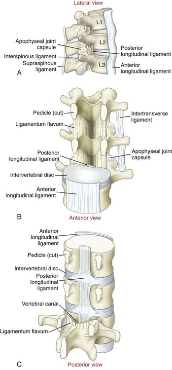

3. Spinal Kinematics: How and Why the Spine Moves
The spine moves in all three cardinal planes: flexion/extension (sagittal), lateral flexion (frontal), and rotation (transverse). However, the amount and primary direction of movement vary significantly between the cervical, thoracic, and lumbar regions. This variation is determined by two key factors:
- Direction of Motion: Dictated by the orientation of the facet (zygapophyseal) joints.
- Amount of Motion: Influenced by the size (height) of the intervertebral disc.
SPINE KINEMATICS(BIOMECHANICS OF SPINE)Physiotherapy Tutorial
This "Movement Science" video provides a brilliant and easy-to-understand explanation of how facet orientation and disc size determine regional spinal movement.
Watch the segment from this point. Focus on how the plane of the facets dictates the primary movement available in each spinal region.
To summarize this crucial concept for your exams:
- Lumbar Spine: Facets are oriented primarily in the sagittal plane. This facilitates flexion and extension while limiting rotation.
- Thoracic Spine: Facets are oriented closer to the frontal plane. This facilitates lateral flexion and rotation. The rib cage provides additional stability, limiting overall motion.
- Cervical Spine: Facets lie in an oblique plane, roughly halfway between the frontal and transverse planes (around 45 degrees). This orientation permits a large range of motion in all three planes.
4. Arthrokinematics: Motion at the Joint Level
Now, let's look at what happens at the joint level during these movements. This is clinically vital because these small motions directly affect the space available for the nerve roots exiting the spine.
SPINE KINEMATICS(BIOMECHANICS OF SPINE)Physiotherapy Tutorial
The same "Movement Science" video continues to break down the specific arthrokinematics of flexion and extension.
Watch the detailed explanation of the mechanics of flexion and extension. Pay close attention to what happens to the intervertebral foramen and the disc.
Here’s a breakdown of the key arthrokinematic events:
During Spinal Flexion (bending forward):
- The superior vertebra tilts and glides anteriorly.
- The facet joints open (or up-glide).
- The intervertebral foramen (IVF), the opening for the nerve root, widens.
- The anterior portion of the annulus fibrosus is compressed, while the posterior portion is tensed.
- Posterior ligaments (PLL, ligamentum flavum, interspinous, supraspinous) become taut.
During Spinal Extension (arching backward):
- The superior vertebra tilts and glides posteriorly.
- The facet joints close (or down-glide).
- The intervertebral foramen (IVF) narrows.
- The posterior portion of the annulus fibrosus is compressed, while the anterior portion is tensed.
- The anterior longitudinal ligament (ALL) becomes taut.
This relationship is the biomechanical basis for many clinical tests and observations. For example, a patient with spinal stenosis (narrowing of the foramen) often finds relief in flexion (which widens the foramen) and experiences increased symptoms in extension (which narrows it).
5. Kinetics: Handling Loads
In our last lesson, we defined torque as Force × Moment Arm. This principle is perfectly illustrated in the biomechanics of lifting.
[PDF] Biomechanics of The Spine | Institute of Motion
This document provides clear diagrams and explanations of how lifting mechanics affect intradiscal pressure.
Read the section on Biomechanics and Lifting. Focus on Figures 14 and 15 and the accompanying text, which explains how lever arms and muscle forces contribute to disc load.
When you lift an object, the external load creates a flexion torque on your spine. Your back extensor muscles must generate an equal and opposite extension torque to maintain equilibrium.
Torque (muscles) = Torque (load)
Because the back muscles have a very short internal moment arm (IMA) compared to the long external moment arm (EMA) of the weight in your hands, they must generate forces many times greater than the weight being lifted. Holding the load closer to your body reduces the EMA, thereby decreasing the required muscle force and the resulting compressive load on the intervertebral discs.
Conclusion
Today, we've dissected the intricate biomechanics of the spinal column, moving from its basic functional unit to the complex interplay of its passive and active components.
Key Takeaways for Your Exam Revision:
- Motion Segment: The functional unit is two vertebrae plus the IVD.
- IVD: The nucleus pulposus handles compression, while the annulus fibrosus resists tension and torsion.
- Ligaments: Know the primary motion each major ligament resists (e.g., ALL resists extension; PLL and posterior ligaments resist flexion).
- Facet Orientation: This is the key determinant of the direction of motion in each spinal region (Lumbar = Flex/Ext; Thoracic = Rot/Lat Flex; Cervical = All-around).
- Arthrokinematics: Flexion opens the facets and widens the IVF; Extension closes the facets and narrows the IVF. This is a critical clinical concept.
- Lifting Kinetics: Keeping a load close to the body minimizes the external moment arm, which reduces the required muscle force and spinal compression.
In our next lesson, we will shift our focus from the axial skeleton to the appendicular skeleton as we describe the biomechanics of the shoulder complex, with a special emphasis on the clinically crucial concept of scapulohumeral rhythm.