Recommended resources
The freely available resources this lesson will direct you to.
- The Lens - Monochromatic Aberrations (Spherical, Coma, Petzval, Distortion) - Episode 3.2www.youtube.com
- Optical aberrations: ray aberrations, wavefront error, Seidel, Abbe sine condition, Zernikewww.youtube.com
- Molecular Expressions Microscopy Primer: Physics of Light and Color - Common Optical Defects in Lens Systems (Aberrations)micro.magnet.fsu.edu
- [PDF] Chapter 20 Aberration Theorywww.erbion.com
Hello. Last lesson treated depth of field as a tolerance on defocus: a scene point had an ideal image location, and we calculated the blur produced when film or sensor lay elsewhere. A real lens presents a different problem. Even when the image plane is placed correctly, rays from one scene point may not agree on where its image belongs.
This lesson gives you a way to recognize six such failures by their physical causes and image signatures. Five occur even with monochromatic light; chromatic aberration requires a range of wavelengths. Together, they explain why photographic lens design could not end with finding the right focal length and aperture.
From a ray miss to an image signature
In paraxial optics, the small-angle approximation makes rays from an object point converge to a single image point. At larger aperture angles or farther off axis, tracing the rays with Snell’s law reveals departures from that prediction. The transverse ray aberration is the distance between a ray’s actual arrival position and its intended position on a chosen image plane. A spot diagram collects those arrival positions for many rays from one object point.
There is a complementary wave description. Ideally, rays converging on an image point belong to a spherical wavefront centered there. An aberrated lens produces an optical-path difference, or wavefront error , across its pupil. Variations in the slope of change ray directions; the resulting image of a point is the diffraction pattern modified by that error. This links the geometric shapes below to the diffraction-limited point image you met earlier, rather than treating them as competing accounts of light.
Optical aberrations: ray aberrations, wavefront error, Seidel, Abbe sine condition, Zernike
Watch the opening and selected passages from Sander Konijnenberg’s “Optical aberrations: ray aberrations, wavefront error, Seidel, Abbe sine condition, Zernike.” They establish the ray and wave descriptions used throughout this lesson.
Watch the setup for the limits of paraxial imaging and the roles of dispersion and construction errors. Then use ray diagnostics to distinguish a ray’s displacement from a spot diagram. Finally, focus and diffraction distinguishes a correctable shift of the image plane from the finite diffraction pattern of a well-focused point.
A useful organizing question is not simply “Is the corner soft?” It is: Does the error depend on position within the aperture, position within the image field, wavelength, or some combination? Those dependencies give the six aberrations their distinct signatures.
Five monochromatic ways to miss the ideal image
For a centered, rotationally symmetric lens, the five primary Seidel aberrations form a compact classification. Let denote a normalized position in the pupil and a normalized position in the image field, measured from its center. After setting aside ordinary focus and magnification, a schematic third-order wavefront expansion is
The coefficients depend on the lens design. The terms, in order, describe spherical aberration, coma, astigmatism, field curvature, and distortion. This is a classification of the leading errors, not a claim that every real lens exhibits each one in isolation.
Read the field dependence first. Spherical aberration can exist at , on the optical axis. Coma increases initially with field position. Astigmatism and field curvature grow with its square, while primary distortion has a cubic field dependence. Now read the pupil dependence: an error that varies across the pupil can spread a point’s rays, but a term linear in shifts them together. That distinction will make distortion unlike the other four.
For the visual counterpart, watch the classification and its spot diagrams:
Optical aberrations: ray aberrations, wavefront error, Seidel, Abbe sine condition, Zernike
Continue Konijnenberg’s video through its five Seidel terms. Watch how each expression changes the image of a point, then how the effect varies across a grid of object points.
Watch the five forms. The wavefront expansion begins around 00:21:42; spot diagrams begin around 00:25:15; the grid of point images begins around 00:29:51. Pay particular attention to why field curvature changes where best focus lies, while distortion changes where an otherwise sharp point lands.
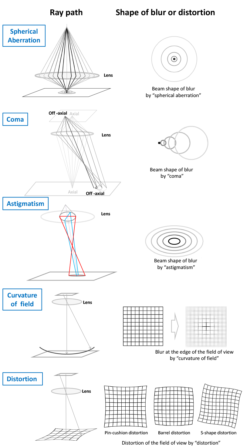
Spherical aberration: pupil zones disagree on axial focus
Consider one on-axis star imaged through a large aperture. Rays passing near the pupil center and rays passing near its rim do not generally cross the axis at the same longitudinal position when the lens’s exact refraction departs from the paraxial prediction. A simple spherical surface often produces this behavior, although its amount and sign depend on surface shape and conjugates; “marginal rays always focus in front” is not a universal rule.
The result is a broadened, approximately rotationally symmetric point image even at field center. Moving the sensor can find a compromise focus, but cannot generally make every pupil zone coincide. Restricting the aperture removes the most extreme zones and often reduces the geometric error substantially. As you saw earlier, however, stopping down also enlarges the diffraction pattern. Spherical aberration is therefore not adequately diagnosed by noting that corners are worse than the center: its defining feature is present on axis.
Coma: an off-axis point grows a tail
For an off-axis point, rays from different pupil zones can produce point images at slightly different transverse positions and scales. Their superposition is asymmetric: a bright concentration with a fan or comet-like tail. In the expansion, coma couples field position to a cubic variation across the pupil, . Its orientation follows the field direction; depending on the sign of the lens’s coma, a tail can point inward or outward.
A photograph of point lights or stars across the frame is especially revealing: the center can look compact while outer points acquire systematically oriented tails. Stopping down commonly reduces coma, but costs light collection. Coma and spherical aberration need not track one another—a design corrected on axis can still image oblique bundles poorly.
Two distinct reasons the edge may not focus
Astigmatism and field curvature can both make the edges of a flat photograph look soft. Their through-focus behavior separates them.
Off-axis astigmatism occurs because ray fans in two perpendicular sections of an oblique bundle do not share the same best-focus position. The tangential section contains the optical axis and the off-axis image point; the sagittal section is perpendicular to it. At one image distance, a point narrows into a line in one orientation. At another distance it narrows into an approximately perpendicular line, with an intermediate, more rounded compromise blur. Thus edge detail can appear directionally smeared, and changing focus exchanges one preferred orientation for the other. In a centered, rotationally symmetric design, this Seidel astigmatism vanishes on axis; element tilt or decentering can additionally create astigmatism where that symmetry no longer holds.
Field curvature means that best focus changes with field position. If sagittal and tangential best-focus locations coincide but trace a curved surface, a flat film or sensor cannot occupy that entire surface. Focus the center and the edges blur; refocus for the edges and the center blurs. Unlike astigmatism, pure field curvature does not require two perpendicular edge focal lines. When astigmatism is present, it is more accurate to speak of separate sagittal and tangential focal surfaces than of one uniquely sharp curved surface.
This is why an edge-sharpness measurement at only one focus setting can mislead. A substantial improvement after refocusing indicates a focus-surface problem; orientation-dependent changes through focus indicate astigmatism. Both effects may coexist.
Distortion: sharp points in the wrong places
Distortion does not principally enlarge the image of a point. It changes the mapping from an object point’s field position to its image position. In the expansion, its wavefront term is linear in pupil position for any fixed : the associated wavefront slope displaces the bundle’s rays together rather than spreading them. The displacement differs across the field, so an image can be sharp yet geometrically inaccurate.
For a simple radial model, let be an ideal image radius and write the actual radius as
With this convention, gives barrel distortion: magnification decreases outward and straight grid lines bow away from the image center. For , pincushion distortion increases magnification outward and the lines bow toward the center. A change of distortion sign with radius can yield a more complex “moustache” pattern. The familiar names describe the grid geometry, not the sharpness of its intersections.
Stop position and multi-element layout influence this mapping. Stopping down may help several blur-producing aberrations, but it generally does not straighten a distorted grid simply by admitting fewer rays. Also distinguish unintended distortion from a deliberately chosen wide-angle projection: not every curved line in a fisheye image represents failure to meet that lens’s design goal.
Color adds a second coordinate: wavelength
The Seidel classification assumed one wavelength. Glass has a refractive index that varies with wavelength, so a refracting element generally has wavelength-dependent optical power. Even if each color were individually well focused, their images need not coincide.
Read the “Chromatic Aberration” portion of the Molecular Expressions Microscopy Primer for its account of dispersion, colored halos, and differences in image magnification.
In the “Chromatic Aberration” subsection, begin with dispersion and focus. After the “Interactive Tutorial” insert, continue with magnification and correction. Focus on the distinction between colors focusing at different axial positions and producing differently sized images. The final discussion of a glass doublet anticipates the next lesson.
Those two effects have different photographic signatures:
- Longitudinal, or axial, chromatic aberration is a wavelength-dependent best-focus distance. It can affect an on-axis point: at one sensor position, one color is more sharply focused while others make colored halos. Changing focus changes which color is sharp and can change the halo’s appearance.
- Lateral, or transverse, chromatic aberration is a wavelength-dependent image magnification. Colors increasingly fail to register as field radius grows. It produces opposed color fringes along high-contrast edges near the frame boundary, with little or no lateral separation at the center of a rotationally symmetric lens. Refocusing alone cannot make the differently sized color images coincide.

Both can coexist. A colored edge by itself therefore does not identify which mechanism dominates: its location across the field and its response to refocusing are more diagnostic than color alone.
Reading an image as an optical test
A practical diagnosis combines three controlled observations. First, compare small point sources at the center and at several field positions. Second, inspect how their shapes change as focus passes through the best setting; where possible, compare a wide aperture with a stopped-down one. Third, include a straight-line grid and, for chromatic effects, high-contrast edges recorded in color. Keep subject distance and image plane position in mind: ordinary subject defocus from the previous lesson can otherwise masquerade as a lens fault.
The expected signatures are compact enough to retain without reducing them all to “softness”:
| Effect | Most telling signature |
|---|---|
| Spherical aberration | Approximately symmetric point blur even on axis; depends strongly on admitted pupil zones |
| Coma | Off-axis points have oriented, asymmetric tails |
| Astigmatism | Off-axis point blur changes between roughly perpendicular line orientations through focus |
| Field curvature | Best focus shifts between center and edge on a flat recording plane |
| Distortion | Grid geometry bends while local points can remain sharp |
| Longitudinal chromatic aberration | Color-dependent focus and halos, possible on axis |
| Lateral chromatic aberration | Color-channel displacement increasing toward the field edge |
These are signatures of idealized, centered aberrations. A real photograph can contain several at once, alongside defocus, diffraction, manufacturing misalignment, and processing artifacts. The classification remains useful because it tells you which comparison to make next, not merely what label to attach to a flawed image.
Conclusion
A paraxial lens has one image position for each scene point; a real lens must also reconcile rays from different pupil positions, field positions, and wavelengths. Spherical aberration separates pupil zones on axis; coma and astigmatism make off-axis points asymmetric or directionally focused; field curvature moves best focus across the frame; distortion moves otherwise sharp points to incorrect positions; and dispersion separates colors in focus, magnification, or both.
These constraints shaped the evolution of photographic optics. In the next lesson, we will examine how achromats, rapid rectilinear lenses, Petzval designs, and anastigmats addressed different combinations of them—and why solving one design problem did not automatically solve the others.