Recommended resources
The freely available resources this lesson will direct you to.
Hello. In the previous lesson, you distinguished plant and animal cells by looking for features such as a cell wall, chloroplasts, and a large central vacuole. Now the question changes from “What is this structure?” to “How does its structure help it do its job?”
This lesson follows the major organelles through the work of a cell: holding instructions, making and processing materials, supplying energy, and storing or breaking down substances. The aim is to make your exam answers more precise than a list of organelle names and functions.
Read a cell diagram as a working system
The labeled comparison below shows a bacterium, a plant cell, and an animal cell. Use the plant and animal cells as a map for this lesson. You will return to several labels that were only briefly discussed last time.
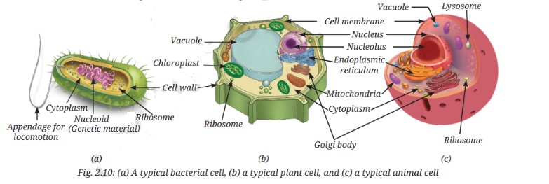
First, watch the selected portion of Biology: Cell Structure from Nucleus Medical Media. Its animation gives you a sense of where structures sit relative to one another. Treat it as an overview; the NCERT readings that follow provide the structural details to use in answers.
Biology: Cell Structure I Nucleus Medical Media
Watch Biology: Cell Structure from Nucleus Medical Media to see the major organelles in the context of whole cells.
Watch the organelle overview, beginning with the nucleus and ending with chloroplasts and the plant cell wall. Notice particularly the free and attached ribosomes, the two kinds of ER, and the movement of materials toward the Golgi apparatus.
An organelle’s function often depends on one of three structural features: a membrane that separates spaces, a large or folded surface where reactions occur, or a particular material inside it, such as DNA, pigment, or enzymes. Keep those three possibilities in mind as you read.
Boundaries make different kinds of work possible
The plasma membrane is the thin outer boundary of every cell. It consists chiefly of a lipid bilayer with proteins embedded in it. The arrangement matters: the water-repelling parts of the lipids face inward, creating a barrier, while membrane proteins can help particular substances cross. Thus the membrane separates the cell from its surroundings without sealing it completely. Its selective permeability will be the focus of the next lesson.
A plant cell also has a cell wall outside that membrane. The wall is relatively rigid and is made primarily of cellulose. Its structure provides support and helps preserve cell shape, but it is permeable; it does not replace the membrane’s selective control over entry and exit.
Read the two short NCERT passages with that contrast in mind.
Read NCERT’s account of the membrane’s lipid-and-protein structure and the plant wall’s supporting role.
On printed pp. 13–14, in “2.2.1 Cell membrane — The universal feature of a cell,” start at the paragraph beginning “Structurally, the cell membrane is” and read the membrane description through the point about proteins helping substances pass. Then, in “2.2.2 Cell wall — The outer covering of cells,” start at “In general, plants cannot move from place to place” and read the wall description through its account of permeability. Focus on why a rigid, permeable wall and a selectively permeable membrane are not interchangeable.
The cytoplasm is the semi-fluid interior in which many cell structures are situated and many reactions take place. In a bacterium, which lacks membrane-bound organelles, much cellular activity occurs directly there. In plant and animal cells, internal membranes create additional spaces with different jobs.
From genetic instructions to usable proteins
Consider how a cell produces a protein that it will release. Its genetic instructions are in the nucleus, whose double-membrane nuclear envelope separates its contents from the cytoplasm. Nuclear pores provide passages for materials moving between the two. Inside are DNA-containing chromatin and the nucleolus, where ribosomal subunits are made. Before cell division, the chromatin becomes organised into visible chromosomes; a non-dividing cell does not usually show the familiar distinct chromosome shapes.
The ribosomal subunits assemble into ribosomes in the cytoplasm. Ribosomes have no surrounding membrane and are sites of protein synthesis. Some are free in the cytoplasm; others attach to the rough endoplasmic reticulum (RER). “Rough” describes the appearance produced by those attached ribosomes. RER is part of a membrane network and is prominent in cells actively making proteins for secretion. Smooth ER (SER) lacks attached ribosomes and is a major site of lipid synthesis.
Materials made in association with the ER can reach the Golgi apparatus in small membrane-bound vesicles. The Golgi consists of stacks of flattened sacs. It modifies, sorts, and packages materials for delivery within the cell or secretion outside it. Some of its packaging forms lysosomes: single-membrane sacs containing digestive enzymes that break down unwanted materials and worn-out cell parts. The membrane keeps this digestive compartment distinct from the surrounding cytoplasm.
Read the NCERT sequence as connected work, not as five unrelated definitions.
Read NCERT’s descriptions of how cellular instructions, synthesis, packaging, and breakdown are distributed among structures.
On printed pp. 16–18, within “2.3.1 Why do eukaryotic cells need these organelles?”, begin under “Nucleus — House of coded instructions” at “The nucleus has a double-layered” and read the organelle sequence through “Lysosomes — The clean-up system,” ending where breakdown products may be reused. Pay particular attention to the “Ribosomes — The protein factories,” “Endoplasmic Reticulum (ER) — Manufacturing factory,” and “Golgi apparatus — The packaging and shipping centres” subsections. You can skip the scientist sidebar; follow instead what each structure is made of or shaped like, and what that enables it to do.
In an exam response, “rough ER makes proteins” needs one more link: ribosomes attached to the RER synthesize the proteins, while the ER provides membrane-enclosed space involved in their processing and transport. Likewise, the Golgi packages and modifies material; it is not the same structure as a ribosome.
The ER, Golgi apparatus, lysosomes, and vacuoles are often grouped as the endomembrane system because their functions are coordinated through membrane-bound compartments. A mitochondrion is also membrane-bound, but it is not part of that particular system.
Two energy-related organelles, two different jobs
Both plant and animal cells need mitochondria. A mitochondrion has an outer membrane and an inner membrane folded into cristae. The folds increase the surface area available for reactions associated with ATP production. Its inner compartment, the matrix, contains components involved in respiration. Mitochondria are sites of aerobic respiration, producing ATP that cells use to power activities. “Powerhouse” is a useful reminder, but folded inner membrane, respiration, ATP is the stronger structural explanation.
A photosynthetic plant cell may also contain chloroplasts, a type of plastid. A chloroplast has a double membrane surrounding the stroma, which contains enzymes involved in making carbohydrates. Inside are flattened membrane sacs called thylakoids, often arranged in stacks called grana. Chlorophyll in the thylakoid membranes absorbs light for photosynthesis. The location of the pigment and the internal membranes help explain why a chloroplast can capture light energy rather than merely appear green.
Read NCERT’s descriptions of these internal structures. You do not need to learn the detailed reaction pathways yet; later modules return to photosynthesis and respiration.
Read the NCERT Biology account for the structural details that connect each organelle to its energy-related role.
In “8.5.4 Mitochondria” on printed p. 97, begin at “Mitochondria (sing.: mitochondrion)” and read the mitochondrion account to the end of that subsection. Then, in “8.5.5 Plastids” on printed pp. 97–98, begin at “The chloroplasts contain chlorophyll and carotenoid pigments” and read the plastid account through the thylakoid description. Focus on cristae versus thylakoids and stroma; leave the detailed reactions for the later photosynthesis and respiration lessons.
Do not treat these organelles as alternatives. Chloroplasts capture light energy to make sugars; mitochondria help release usable energy from food during respiration. Plant cells respire too. The plastid family also includes pigment-containing chromoplasts, which contribute colours to some flowers and fruits, and colourless leucoplasts, which store food materials.
Both mitochondria and chloroplasts contain some of their own DNA and ribosomes and can make some of their own proteins. That is a useful structural fact, but it does not mean either organelle can function independently as a complete cell.
Storage, support, and movement
In a typical mature plant cell, the large central vacuole is a single-membrane-bound compartment filled with watery cell sap. It stores substances including water, minerals, sugars, and wastes. Storing water also helps maintain pressure inside the cell; together with the cell wall, this helps keep the cell firm. When water is lost, that support decreases and the plant may wilt. Animal cells can have vacuoles, but they usually do not have the same large central one.
Read NCERT’s concise account of how the plant vacuole’s contents contribute to storage and firmness.
On printed p. 20, under “Vacuoles — The organelles for storage and support,” start at the sentence beginning “In a mature plant cell, there is usually one” and read the vacuole account through the comparison with animal cells. Notice the single surrounding membrane, cell sap, and the consequence of losing stored water.
Not all support comes from walls or fluid pressure. The cytoskeleton is a network of protein fibres in the cytoplasm that helps maintain shape, organise internal structures, and enable movement. Some animal cells also have a centrosome containing centrioles, structures involved in organising the spindle used during cell division. You will revisit the spindle when you study mitosis and meiosis.
Cilia and flagella are projections that help move a cell or fluid near its surface. Their structure matters too: eukaryotic cilia and flagella contain organised microtubules. The bacterium’s locomotory appendage in the opening diagram should not be assumed to have that same internal structure.
Key takeaways
To relate an organelle’s structure to its function, name the feature that makes the function possible:
- Nuclear pores permit exchange with the DNA-containing nucleus; ribosomes provide sites for protein synthesis.
- Ribosome-studded RER, smooth ER, and flattened Golgi sacs have different roles in making, handling, and packaging materials.
- Enzymes within membrane-bound lysosomes break down unwanted material.
- Mitochondrial cristae provide increased membrane area for processes that produce ATP; chlorophyll-bearing thylakoids capture light for photosynthesis.
- A fluid-filled central vacuole stores materials and helps maintain plant-cell firmness.
The cell works through cooperation among these structures, not through any one organelle acting alone. Next, you will examine one important part of that cooperation in detail: how substances move across the plasma membrane, and when that movement does or does not require cellular energy.