Recommended resources
The freely available resources this lesson will direct you to.
Hello. Last lesson, you used the presence of a nucleus and membrane-bound organelles to distinguish eukaryotic cells from prokaryotic cells. Plant and animal cells both meet those eukaryotic criteria, so today you need a closer look: which visible structures distinguish one from the other?
This lesson focuses on identifying evidence in diagrams and microscope observations, then turning that evidence into a precise exam answer. The detailed functions of organelles come next; for now, a few functions will help you remember what you are seeing.
Begin at the cell’s boundary
Compare the two schematic cells below. The plant cell has a cell wall outside its cell membrane. The animal cell has a cell membrane but no cell wall. In the drawing, the wall gives the plant cell a more regular outline, while the animal cell has a more flexible boundary.
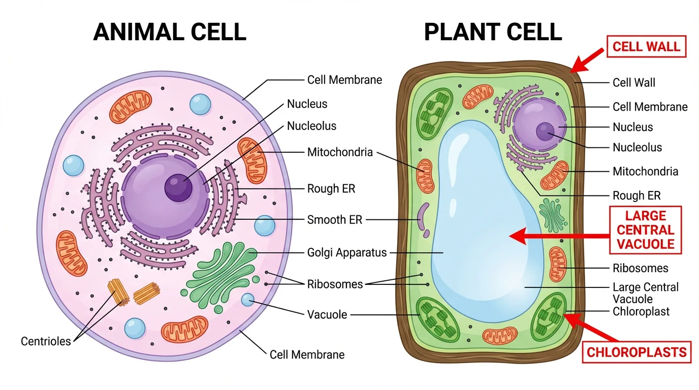
The wall and membrane are two different layers, not two names for the same layer. The wall is a relatively rigid, generally permeable covering; the membrane beneath it controls movement into and out of the cell. An animal cell’s outer boundary is its membrane.
Now connect the drawing to cells viewed under a microscope. Read the short observations in the NCERT chapter Cell: The Building Block of Life.
Read NCERT’s comparison of onion-peel and cheek cells to see which differences are observable, rather than only drawn in a schematic.
In Activity 2.3 on p. 13, find the paragraph beginning with the observations of onion-peel cells. Read the comparison and look at Fig. 2.8. Then, in Section 2.2.2, “Cell wall — The outer covering of cells,” on pp. 13–14, read from the explanation beginning “In general, plants cannot move from place to place” through the comparison with animal cells: the wall explanation. Focus on what keeps the plant cell’s outer shape when its contents shrink.
NCERT describes onion-peel cells as box-shaped and regularly arranged, whereas cheek cells are irregularly arranged. Shape is a clue, not an absolute test: cells take many forms in different tissues. The stronger observation is the plant cell’s rigid wall. NCERT’s concentrated-sugar observation makes the distinction especially clear: the plant cell’s contents can pull away from its wall while the outer wall retains its shape; a cheek cell, lacking that wall, shrinks as a whole. You will study the movement of water across membranes in a later lesson.
Look inside: strong clues and shared structures
Return to the labeled comparison. The plant drawing contains green, oval chloroplasts and a large central vacuole occupying much of the cell. The animal drawing has neither chloroplasts nor a single comparably large central vacuole. It depicts small vacuoles instead.
Watch these selected parts of Plant Cell vs. Animal Cell from Henrik’s Lab for a moving comparison of the wall, chloroplasts, vacuole, and an animal-cell feature.
Watch these portions for the visual contrast between a plant cell’s prominent structures and those commonly drawn in an animal cell.
Start with the outer wall, noticing its position outside the membrane. Then watch chloroplasts and mitochondria: unlike chloroplasts, mitochondria occur in both cells. Continue with the central vacuole and the animal centrosome. Keep the comparison specifically between plant and animal cells: a cell wall is not exclusive to plants among all organisms.
The chloroplast contains the green pigment chlorophyll and is associated with photosynthesis. It is an excellent clue when it appears in a plant-cell diagram, but do not demand that every plant cell show one. Onion-peel cells, for example, may not display visible chloroplasts; a plant’s non-green cells need not have them. Conversely, chloroplasts also occur in some organisms other than plants, so context still matters.
The large central vacuole is another strong clue for a typical mature plant cell. Animal cells may have small vacuoles, so “plants have vacuoles; animals have none” would be an inaccurate comparison. The useful distinction is chiefly size and prominence.
NCERT gives the context behind both features. Its discussion also introduces plastids—the broader group to which chloroplasts belong—so you need not mistake “plastid” for a synonym of “green chloroplast.”
Read NCERT’s descriptions to understand why chloroplasts and a large central vacuole are useful, but not universal, visual clues.
On pp. 18–19, under “Plastids — Centre for food synthesis in the plant cells and beyond,” read the plastid description. In the same section, find the paragraph beginning “Some plastids lack pigments” and read the colourless-plastid example. Then, on pp. 19–20, under “Vacuoles — The organelles for storage and support,” read the vacuole comparison. Note NCERT’s qualified wording: “usually” for a mature plant cell and “sometimes” for animal-cell vacuoles.
The vacuole’s stored water helps keep a mature plant cell firm. That explains why a large clear region can dominate a plant-cell drawing, with other structures occupying the space around it. A schematic may show that arrangement plainly; an ordinary classroom microscope image may reveal fewer details. Do not treat every labeled structure in a textbook schematic as something you must be able to see in an onion-peel slide.
Decide what counts as evidence
Because both cells are eukaryotic, several conspicuous parts do not distinguish them. Both can contain a nucleus, cell membrane, cytoplasm, mitochondria, ribosomes, endoplasmic reticulum, and Golgi apparatus. A nucleus tells you the cell is eukaryotic; by itself, it does not tell you whether it is plant or animal.
| Feature in a typical diagram | Plant cell | Animal cell |
|---|---|---|
| Cell wall outside cell membrane | Present | Absent |
| Chloroplasts | Present in photosynthetic cells | Absent |
| Vacuole | Usually one large central vacuole in a mature cell | May have smaller vacuoles |
| Cell membrane and nucleus | Present | Present |
| Mitochondria, ribosomes, ER, Golgi apparatus | Present | Present |
Some animal-cell diagrams also label centrioles, as the supplied comparison does. Their depiction may support an animal-cell identification, but the wall, chloroplasts, and large central vacuole provide the more useful introductory comparison. Likewise, a roughly round outline is weaker evidence than a labeled structure.
One qualification carries forward from the previous lesson: a wall alone does not prove that a cell is a plant cell. Bacteria and fungi can have walls too. If a question asks you to choose only between a plant cell and an animal cell, a wall distinguishes the two. If other kinds of cells are possible, inspect additional evidence, particularly whether there is a well-defined nucleus and whether plant-associated structures are shown.
See how NCERT’s exemplar questions use shared structures as distractors.
Chapter 8 cell-structure and function.pmd
Inspect a few exam-style options to see why a familiar organelle is not necessarily a distinguishing feature.
Under “MULTIPLE CHOICE QUESTIONS,” find Question 3 on p. 40 and read its combinations. Then go to Questions 18–20 on p. 42, beginning with the question about distinguishing a plant cell from an animal cell, and read the three questions. Notice how Question 19 tests the mistake of equating any cell wall with a plant cell.
For a short diagram-based answer, name an observation and state the inference it supports. For example: “The cell has a wall outside its membrane and a large central vacuole; these features identify it as a plant cell rather than an animal cell.” If chloroplasts are shown, they provide another supporting observation. Avoid an answer such as “It is a plant cell because it has a nucleus”: the nucleus is shared.
Key takeaways
To distinguish plant and animal cells, start with the outer boundary, then check for chloroplasts and the size of the vacuole. A typical plant-cell diagram shows a wall, and often chloroplasts and a large central vacuole; an animal cell lacks the wall and chloroplasts and does not have that characteristic large central vacuole. The nucleus and many other organelles are shared, not distinguishing features. Use shape as a supporting clue, and allow for differences between cell types and between schematics and microscope views.
Next, you will examine the major organelles more closely and relate each organelle’s structure to its function.