Recommended resources
The freely available resources this lesson will direct you to.
Hello. In the previous lesson, you identified the building units of carbohydrates, proteins, and nucleic acids, and saw why lipids need a different description. Now the question is more precise: what joins those units together?
This lesson focuses on four bond names that are easy to confuse in exams: glycosidic, peptide, phosphodiester, and ester. By the end, you should be able to recognise each from the molecules it connects and, where a diagram is given, from the part of the structure that forms the link.
A bond has a name because of what it connects
When small biological molecules join, they form a covalent bond: the connected atoms share electrons. Often, forming the link releases water in a condensation or dehydration reaction. Hydrolysis is the reverse kind of reaction: water is used to split a link. These are useful patterns, but “covalent bond” and “dehydration” are not specific enough when an exam asks you to name the bond.
Watch Dehydration Synthesis and Hydrolysis by Amoeba Sisters for the visual distinction between forming a link and breaking one. Pay particular attention to the different names given to the links in proteins and carbohydrates.
Dehydration Synthesis and Hydrolysis
Watch Dehydration Synthesis and Hydrolysis by Amoeba Sisters to see how removing water can join building units and how adding water can separate them.
Begin with the joining idea, then watch amino acids to locate the peptide bond. Continue with two sugars to identify a glycosidic bond. Finish with hydrolysis and notice that it breaks an existing linkage rather than naming a new type of bond.
Keep the distinction between process and product clear. Condensation describes a way a link is made; a peptide bond, for example, names the particular link left between two amino acids.
Glycosidic bonds join sugars; ester bonds attach fatty acids
A monosaccharide such as glucose can join another monosaccharide through a glycosidic bond (also called a glycosidic linkage). Two joined sugar units form a disaccharide; many joined units form a polysaccharide. Thus, starch, glycogen, and cellulose contain glycosidic linkages between their sugar units. The name of the linkage alone does not tell you whether the resulting carbohydrate stores energy or provides structure.
Lipids require different wording. In a triglyceride, three fatty acids attach to one glycerol molecule by ester bonds. Fatty acids are not repeating monomers in a long triglyceride chain: the ester bonds attach them to a glycerol backbone.
Read the two short parts of NCERT’s Biotechnology biomolecules chapter below. They put the bond names alongside the molecules they connect.
Read NCERT’s account of sugar linkages and fatty-acid attachment to glycerol to fix the two bond names in context.
On p. 52, find “2. Oligosaccharides” and read the explanation of joined sugars. Immediately below, in “3. Polysaccharides,” read the opening account, through its examples. Then go to “3.2 Fatty Acids and Lipids” on p. 60. From the opening sentence about lipids in living organisms, read the introductory paragraph. Focus on what the ester linkage connects, rather than on the later classification of lipid types.
The ester bonds are visible in this reaction diagram. Each circled region on the product side contains a link between glycerol’s oxygen and a fatty acid’s carbonyl carbon; forming the triglyceride releases three water molecules.

There is a useful visual warning here. A glycosidic linkage and an ester linkage both contain oxygen, but an oxygen atom by itself does not identify the bond. Look at the groups on either side: sugar joined to sugar suggests glycosidic; fatty acid joined to glycerol suggests ester.
Peptide bonds join amino acids; phosphodiester bonds join nucleotides
An amino acid has an amino group and a carboxyl group. When the carboxyl group of one amino acid links to the amino group of another, the resulting connection is a peptide bond. Repeating that connection produces a polypeptide chain. In a structural formula, the characteristic portion of the link is : the bond joins the carbonyl carbon of one amino-acid residue to the nitrogen of the next.
DNA and RNA are chains of nucleotides, not chains of nitrogenous bases alone. Each nucleotide contains a sugar, a phosphate group, and a base. Adjacent nucleotides in a strand are joined by a phosphodiester linkage: a phosphate connects the sugars of neighbouring nucleotides. More precisely, it links the position of one sugar to the position of the next, producing the repeating sugar–phosphate backbone.
Read NCERT’s descriptions of these two chains. You do not need to memorise every structural detail in the chapter to identify the bonds; concentrate on the groups joined and the resulting backbone.
Return to NCERT’s Biotechnology biomolecules chapter for its descriptions of peptide and phosphodiester linkages.
In “3.4.1 Primary Structure of Proteins” on pp. 68–69, start at the description of a linear amino-acid chain and read the bond explanation, including Figure 3.24. Note which group each amino acid contributes. In “3.5 Nucleic Acids” on p. 76, read the opening description. Then locate “3.5.1 Polynucleotide Chain” on p. 79 and read the linkage description, using Figure 3.32 to locate the phosphate between two sugars.
For a brief visual check of the last link, watch this excerpt from Biological Molecules: Carbohydrates, Lipids, Proteins & Nucleic Acids by EKG Science.
Biological Molecules: Carbohydrates, Lipids, Proteins & Nucleic Acids
Watch the nucleotide-chain portion of EKG Science’s Biological Molecules: Carbohydrates, Lipids, Proteins & Nucleic Acids to see why the link belongs to the sugar–phosphate backbone.
Watch the nucleotide link. Follow the phosphate bridge between neighbouring sugars; the bases are attached to the sugars but are not the links joining successive nucleotides in a strand.
One distinction matters especially in DNA diagrams. Hydrogen bonds between complementary bases hold the two DNA strands together. They are not the phosphodiester bonds that join successive nucleotides within each strand. Identify which connection a question points to before naming it.
Reading an unfamiliar diagram
The four rows of this overview show how small components relate to larger biological structures. Use the labelled bonds in the chemical drawings on the left; the folded protein and DNA helix on the right show larger structures, not additional names for the links between successive building units.
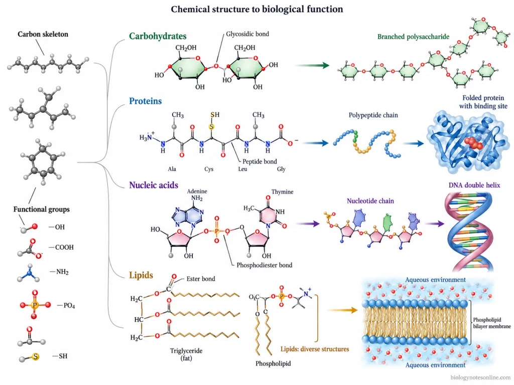
For a short exam answer, pairing what joins with the bond name is usually more reliable than listing bond names alone:
| Connection shown or described | Bond to identify |
|---|---|
| One sugar unit joined to another | Glycosidic bond |
| One amino acid joined to the next in a polypeptide | Peptide bond |
| A phosphate bridging successive sugars in a DNA or RNA strand | Phosphodiester bond |
| A fatty acid attached to glycerol in a fat | Ester bond |
The last row is deliberately phrased differently. Proteins, nucleic acids, and polysaccharides are polymers with links between successive building units; a triglyceride is not a polymer of repeating fatty-acid monomers. Ester is still the correct name for its fatty acid–glycerol attachments.
Key takeaways
Name a linkage by locating the two components it connects: sugars use glycosidic bonds, amino acids use peptide bonds, and nucleotides in a strand use phosphodiester bonds. In common fats, fatty acids attach to glycerol by ester bonds, without making the fat a repeating-unit polymer. Condensation and hydrolysis describe joining and splitting reactions; they do not replace those specific bond names.
In the next lesson, the focus moves from molecular building and linking to enzymes—molecules that catalyse reactions—and how temperature, pH, and substrate concentration affect their action.