Welcome to your final lesson on joints and articulations. In our previous session, we examined the shoulder, a joint designed for maximum mobility. Now, we will turn our attention to the knee, the largest and one of the most complex joints in the body, which, by contrast, is built primarily for stability and weight-bearing.
Your goal for this lesson is to describe the key structural features and major movements of the knee joint. For a nursing student, a solid understanding of the knee is essential. You will frequently encounter patients with knee injuries—from athletic tears to osteoarthritis, which is common in the older adult population you've worked with. This knowledge will be invaluable for patient assessment, understanding treatment plans, and providing effective education.
We will deconstruct the knee by examining its bony framework, the crucial soft tissues that provide stability (like ligaments and menisci), and the primary movements it allows.
Articulating Bones and Surfaces
The knee joint is a modified hinge-type synovial joint. It is formed by three bones: the femur (thigh bone), the tibia (shin bone), and the patella (kneecap). It's important to note that the fibula, the other bone in the lower leg, does not participate in the knee joint itself.
These bones form two distinct articulations within a single joint capsule:
- Tibiofemoral joint: The articulation between the large condyles of the femur and the relatively flat plateaus of the tibia. This is the primary weight-bearing component of the knee.
- Patellofemoral joint: The articulation between the patella and a groove on the anterior surface of the femur.
Let's begin with a video that clearly introduces these foundational components.
Knee Joint Anatomy & Common Injuries (Ligaments, Menisci & Movements)
This video from Taim Talks Med provides a clear, animated breakdown of the bones and articulating surfaces that make up the knee joint.
Watch the segment from bones and articulations. Pay close attention to the three bones involved and how they form the two distinct joints (tibiofemoral and patellofemoral).
Key Stabilizing Structures
The stability of the knee does not come from a deep, interlocking bony fit like the hip. Instead, it relies on an intricate network of soft tissues: the menisci, which improve the joint's congruency, and an extensive system of strong ligaments.
Menisci: The Shock Absorbers
Sitting on the tibial plateaus are two C-shaped pads of fibrocartilage called the menisci (singular: meniscus). You can think of them as gaskets that serve two critical functions:
- They act as shock absorbers, cushioning the joint and distributing body weight evenly across the tibia.
- They deepen the tibial plateaus, creating a more stable "cup" for the femoral condyles to sit in, much like the glenoid labrum does in the shoulder.
There are two menisci: the medial meniscus (C-shaped) on the inner side of the knee and the lateral meniscus (more circular) on the outer side.
Ligaments: The Joint's Ropes
The ligaments of the knee are tough, fibrous bands that connect the bones and prevent excessive or abnormal movement. They are the primary source of the knee's stability and can be grouped into two main categories.
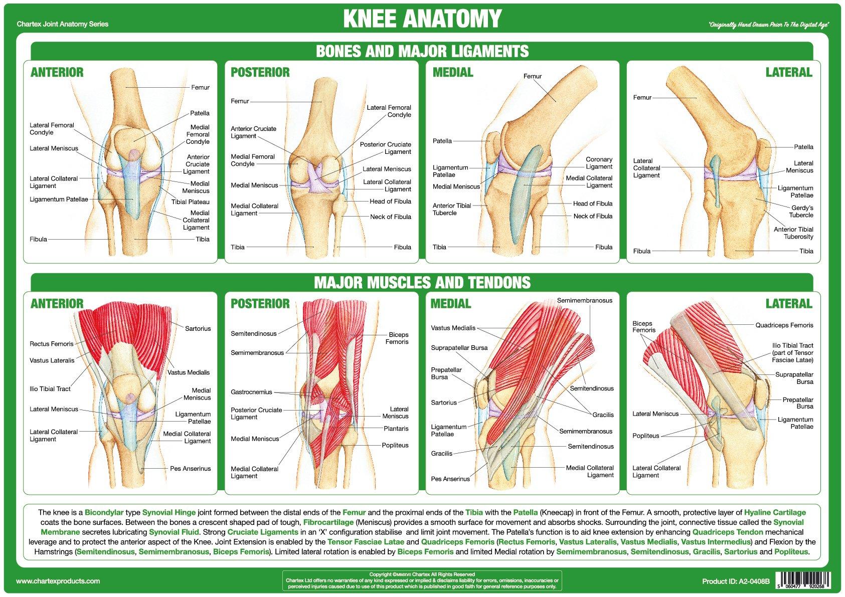
1. Extracapsular Ligaments (Outside the Joint Capsule)
- Tibial (Medial) Collateral Ligament (MCL): Runs along the inner side of the knee, preventing the knee from buckling outwards (valgus stress).
- Fibular (Lateral) Collateral Ligament (LCL): Runs along the outer side of the knee, preventing the knee from buckling inwards (varus stress).
- Patellar Ligament: Connects the patella to the tibial tuberosity. It is functionally a continuation of the quadriceps femoris tendon.
2. Intracapsular Ligaments (Inside the Joint Capsule)
These two crucial ligaments cross each other inside the joint, forming an "X" shape, which is why they are called "cruciate" ligaments (from the Latin crux, meaning cross).
- Anterior Cruciate Ligament (ACL): Prevents the tibia from sliding too far forward relative to the femur and prevents hyperextension.
- Posterior Cruciate Ligament (PCL): Prevents the tibia from sliding too far backward relative to the femur.
The following reading provides a detailed look at these structures.
Knee joint: anatomy, ligaments and movements
This Kenhub article offers a detailed written description of all the stabilizing structures we are discussing. Use it to solidify your understanding of their locations and functions.
Read the section titled Ligaments and menisci. Start by reviewing the descriptions for the following key structures: Lateral (fibular) collateral ligament Medial (tibial) collateral ligament Cruciate ligaments Menisci As you read, focus on the primary function of each structure—what specific movement does it prevent?

Bursae
Like other complex synovial joints, the knee is surrounded by several bursae. These are small, fluid-filled sacs that act as cushions to reduce friction between bones, tendons, and muscles. Inflammation of a bursa is called bursitis, a common condition you may see in clinical practice (e.g., "housemaid's knee" is prepatellar bursitis).
Movements of the Knee Joint
As a modified hinge joint, the knee's primary movements are flexion and extension. However, it also allows for a small degree of rotation, which is critical for its function.
- Flexion: Bending the knee (e.g., bringing your heel towards your buttock).
- Extension: Straightening the knee.
- Medial & Lateral Rotation: A small amount of inward and outward rotation of the tibia is possible, but only when the knee is flexed.
A unique feature of the knee is the "screw-home mechanism." As the knee moves into full extension, the femur medially rotates slightly on the tibia. This rotation locks the knee into its most stable position, allowing us to stand for long periods without fatiguing our muscles. To initiate flexion from this locked position, the knee must first be "unlocked" by a muscle that causes slight lateral rotation of the femur.
This next video provides an excellent visual explanation of these movements, including the screw-home mechanism.
Understanding the Anatomy of Your Knee
This animation from Muscle & Motion clearly demonstrates the movements of the knee and provides a simple explanation of the crucial screw-home mechanism.
Watch the segment on movement, from movement and the screw-home mechanism. Focus on understanding how rotation "locks" the knee into a stable, extended position.
Conclusion
In this lesson, we have dissected the knee, a joint masterpiece engineered for stability under immense load. Unlike the highly mobile shoulder, the knee's structure prioritizes support and controlled movement.
Key Takeaways:
- The knee is a modified hinge joint consisting of the tibiofemoral and patellofemoral articulations.
- Its stability relies heavily on soft tissues: the menisci provide shock absorption and congruency, while a robust network of ligaments provides reinforcement.
- The key ligaments include the collaterals (MCL and LCL), which prevent side-to-side motion, and the cruciates (ACL and PCL), which prevent forward/backward displacement.
- The primary movements are flexion and extension, with limited rotation possible only when the knee is flexed.
- The screw-home mechanism medially rotates the femur during final extension to "lock" the knee, providing maximum stability for standing.
This lesson concludes our module on joints and articulations. You have now examined the body's skeletal framework and the diverse ways our bones connect to permit or restrict movement. In our next module, we will shift our focus to the engine of movement itself: muscle tissue. We will begin by exploring its structure and the fascinating physiological process that allows it to contract.