Welcome to the first lesson of our module on Muscle Tissue and Physiology. In our last module, we explored how joints create connections between bones to allow for movement. Now, we'll dive into the very tissue that powers that movement. This lesson will provide a foundational overview by comparing the three distinct types of muscle found in the human body.
Your goal is to be able to compare skeletal, cardiac, and smooth muscle tissue based on their structure, location, and how they are controlled. As a future nurse, this knowledge is fundamental. It will help you understand everything from a patient's mobility (skeletal muscle), to their cardiovascular status (cardiac muscle), to the functioning of their digestive and urinary systems (smooth muscle). Your experience in a retirement home has likely shown you the critical importance of muscle function for overall health and quality of life.
We will begin by exploring the key characteristics that differentiate these three muscle types, focusing on their microscopic appearance, where they are in the body, and whether we control them consciously.
An Introduction to Muscle Tissue Types
All muscle tissues share a key property: excitability. Like nervous tissue, they can respond to a stimulus. For muscle, this response is contraction, which generates force and produces movement. However, the way they are structured and controlled leads to three very different roles in the body.
The following video from Dr. Matt & Dr. Mike offers a comprehensive and engaging overview of the three muscle types, covering all the core concepts for this lesson in a single, coherent narrative.
Muscle Tissue | Skeletal, Cardiac, and Smooth
This video provides a detailed comparison of skeletal, cardiac, and smooth muscle tissue. It covers their control, cellular structure, appearance, and how the cells are connected to one another to achieve their specific functions.
As you watch, focus on building a mental table comparing the three types across several key features. First, watch from the beginning to the overview of control. Note the primary function of each muscle type and whether it is under voluntary (conscious) or involuntary (automatic) control. Next, continue from cell shape and nuclei. Pay attention to the differences in cell shape (cylinder, branched, or spindle) and the number and location of nuclei within each cell. Then, watch the section on striations and internal structure. Understand what striations are and why smooth muscle lacks them. Finally, view the segment on cell connections. Note the special structures in cardiac muscle, the intercalated discs, and why they are so important for the heart's function.
Consolidating the Comparison
The video you just watched laid out the essential differences between the three muscle tissues. Let's systematically review and organize these key distinguishing features.
1. Control
This is perhaps the most straightforward distinction.
- Skeletal Muscle: The only type under voluntary control, governed by the somatic nervous system. You consciously decide to move these muscles.
- Cardiac and Smooth Muscle: Both are involuntary, regulated by the autonomic nervous system. You do not consciously think to make your heart beat or your stomach churn.
2. Location
Their location is directly tied to their function.
- Skeletal Muscle: As the name implies, it's primarily attached to the skeleton, enabling body movement, posture, and facial expressions.
- Cardiac Muscle: Found exclusively in the wall of the heart. Its function is singular and vital: to pump blood.
- Smooth Muscle: Located in the walls of hollow organs (like the stomach, intestines, and bladder), airways, and blood vessels. It moves substances through internal passageways.
3. Structure (Microscopic Anatomy)
This is where the differences are most numerous.
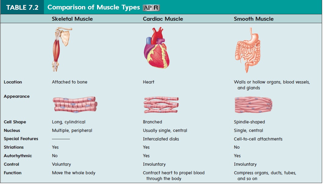
| Feature | Skeletal Muscle | Cardiac Muscle | Smooth Muscle |
|---|---|---|---|
| Cell Shape | Long, cylindrical fibers | Short, branched cells | Spindle-shaped (fusiform) cells |
| Striations | Yes (striped appearance) | Yes (striped appearance) | No (smooth appearance) |
| Nuclei | Multinucleated (many nuclei), located peripherally | Usually uninucleated (one nucleus), sometimes two, located centrally | Uninucleated (one nucleus), located centrally |
| Special Features | - | Intercalated discs containing gap junctions and desmosomes | - |
The striations visible in skeletal and cardiac muscle are due to the highly organized, repeating arrangement of contractile proteins (actin and myosin) into units called sarcomeres. We will explore sarcomeres in detail in a future lesson. In smooth muscle, these proteins are present but arranged less uniformly, hence the "smooth" appearance.
The intercalated discs in cardiac muscle are a crucial feature. These specialized junctions hold the cardiac cells tightly together and contain gap junctions. These are like tiny tunnels that allow electrical signals to pass rapidly from cell to cell. This ensures that all the heart muscle cells contract in a coordinated wave, creating an effective pump—a phenomenon known as a functional syncytium.
The following reading from Registered Nurse RN provides another excellent summary and includes a helpful mnemonic for remembering the locations of smooth muscle.
Three Types of Muscle Tissue: Skeletal, Cardiac, and Smooth Muscle
This article breaks down the characteristics of each muscle type, reinforcing the comparisons we've discussed. It's written from a nursing perspective and offers practical memory aids.
Read the main sections for each muscle type to solidify your understanding: Skeletal Muscle Tissue Cardiac Muscle Tissue Smooth Muscle Tissue Pay special attention to the mnemonic "STOVE" provided in the section on smooth muscle location. This is a great tool for exam preparation.
To bring all of these points together visually, the chart below provides a clear, side-by-side comparison of the three muscle types, summarizing their key features, functions, and appearance.
Conclusion
In this lesson, we established the fundamental distinctions between the three types of muscle tissue. Understanding these differences is the first step toward appreciating the specialized roles muscle plays throughout the body, from powerful, voluntary movements to the subtle, automatic work of our internal organs.
Key Takeaways:
- Skeletal Muscle: Voluntary, striated, multinucleated, and attached to bones for movement.
- Cardiac Muscle: Involuntary, striated, and features branched, single-nucleus cells connected by intercalated discs to form the contractile walls of the heart.
- Smooth Muscle: Involuntary, non-striated, and consists of single-nucleus, spindle-shaped cells found in the walls of hollow organs and vessels.
Having established this broad overview, our next lesson will begin a deeper dive into the most intricate of the three: skeletal muscle. We will explore its complex structural hierarchy, from the entire muscle organ down to the individual protein filaments that make contraction possible.