Welcome to your next lesson on articulations. In our last session, we finished building your vocabulary for describing body movements. Now, we'll apply those concepts by examining the detailed anatomy of one of the body's most complex and important joints: the shoulder.
Your goal for this lesson is to describe the key structural features and major movements of the shoulder joint. As a nursing student, you'll find this knowledge immediately applicable. Shoulder injuries, such as dislocations and rotator cuff tears, are common, and understanding the joint's anatomy is crucial for assessing patients and understanding their treatment and recovery.
We will explore the bones that form the joint, the ligaments that stabilize it, and the critical muscles that both move and protect it.
The Shoulder Joint: A Trade-off Between Mobility and Stability
The shoulder joint, anatomically known as the glenohumeral joint, is a ball-and-socket synovial joint. It's formed by the articulation of the head of the humerus (the "ball") with the glenoid fossa of the scapula (the "socket").
This joint is the most mobile in the entire human body, allowing for an incredible range of motion. However, this flexibility comes at a price: inherent instability. The "socket" is quite shallow, meaning the bones themselves provide very little support. This relationship is often compared to a golf ball resting on a tee.
To get a better sense of this fundamental trade-off, let's begin with a short introductory video.
Glenohumeral joint: Structure and actions
This clip from "Glenohumeral joint: Structure and actions" by The Noted Anatomist introduces the joint's basic structure and explains the relationship between its high flexibility and low stability.
Watch the first two minutes of the video, from the beginning to the introduction. Pay close attention to the "golf ball on a tee" analogy.
Key Structural Features
Because the bony fit of the shoulder joint is so poor, it relies heavily on soft-tissue structures for stability. These include cartilage, ligaments, and a specialized group of muscles.
Articulating Surfaces and the Glenoid Labrum
As you just saw, the main articulating surfaces are the head of the humerus and the glenoid fossa of the scapula. To improve the fit and add some much-needed depth to the shallow glenoid fossa, a fibrocartilaginous ring called the glenoid labrum is attached to its outer rim. This structure acts like a rubber gasket, deepening the socket by about 50% and creating a mild suction effect that helps hold the humeral head in place.
For a clear written explanation of these surfaces, please read the following section.
Glenohumeral (Shoulder) joint: Bones, movements, muscles
This section from a Kenhub article describes the articulating bones and the function of the glenoid labrum.
Please read the section titled Articular surfaces, starting from the description of the joint. Focus on how the glenoid labrum increases the congruency of the joint.
The Joint Capsule and Supporting Ligaments
Like all synovial joints, the shoulder is enclosed in a fibrous joint capsule. This capsule is notably loose or lax, which is necessary to permit the joint's wide range of motion. Consequently, it provides minimal stability on its own and must be reinforced by ligaments and muscles.
Several key ligaments strengthen the joint capsule and prevent excessive movement:
- Glenohumeral Ligaments: A group of three ligaments (superior, middle, and inferior) that reinforce the anterior part of the capsule. They are the primary source of stability and work to prevent anterior dislocation.
- Coracohumeral Ligament: A strong ligament that runs from the coracoid process of the scapula to the humerus, supporting the superior part of the joint.
- Transverse Humeral Ligament: This ligament spans the distance between the two tubercles of the humerus, holding the tendon of the long head of the biceps brachii muscle in place.
The following image provides a detailed view of these ligaments and their relationship to the bones of the shoulder.

To understand how these ligaments are named and how they function together, the next video provides an excellent, easy-to-follow explanation.
Anatomy of the Shoulder Joint | Bones, Ligaments, and Muscles
This video from Siebert Science, "Anatomy of the Shoulder Joint," clearly explains the ligaments and their naming conventions, which makes them much easier to remember.
Watch the portion of the video from the ligaments section. The speaker does a great job of breaking down the complex names into the parts they connect (e.g., "acromio-clavicular").
The Rotator Cuff Muscles
While ligaments provide passive stability, the most important dynamic stabilizers of the shoulder are a group of four muscles known as the rotator cuff. These muscles originate on the scapula, and their tendons wrap around the head of the humerus, forming a "cuff" that pulls the humeral head firmly into the glenoid fossa. This action, called concavity compression, is essential for keeping the shoulder stable during movement.
The four rotator cuff muscles are easily remembered with the mnemonic SITS:
- Supraspinatus
- Infraspinatus
- Teres minor
- Subscapularis
The next segment of the Siebert Science video provides a fantastic overview of these four muscles, their locations, and their functions.
Anatomy of the Shoulder Joint | Bones, Ligaments, and Muscles
This section focuses on the four rotator cuff muscles, explaining where they are and what they do.
Please watch from the rotator cuff section. Focus on the collective function of the SITS muscles in stabilization (concavity compression) and their individual roles in movement.
Major Movements of the Shoulder Joint
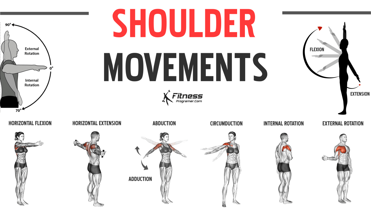
Thanks to its ball-and-socket structure, the shoulder joint allows for movement in all three anatomical planes. You'll recognize these from our previous lessons:
- Flexion and Extension: Moving the arm forward and backward.
- Abduction and Adduction: Moving the arm away from and toward the midline.
- Internal (Medial) and External (Lateral) Rotation: Rotating the arm inward and outward.
- Circumduction: A combination of all the above movements, creating a cone-like motion.
The image below illustrates these movements.
For a dynamic demonstration, let's return to The Noted Anatomist for a final short clip.
Glenohumeral joint: Structure and actions
This brief clip clearly demonstrates each of the primary movements of the glenohumeral joint.
Watch the segment from the actions section to see these movements in action.
Conclusion
In this lesson, we dissected the complex anatomy of the glenohumeral joint. We saw how its structure is a masterclass in compromise, sacrificing bony stability for an unparalleled range of motion.
Key Takeaways:
- The shoulder is a ball-and-socket joint formed by the head of the humerus and the shallow glenoid fossa of the scapula.
- Its inherent instability is counteracted by soft-tissue structures: the glenoid labrum (which deepens the socket), the joint capsule and its reinforcing ligaments (glenohumeral, coracohumeral), and most importantly, the four rotator cuff (SITS) muscles.
- The rotator cuff muscles provide dynamic stability through concavity compression, pulling the humeral head into the glenoid fossa.
- The primary movements of the shoulder are flexion/extension, abduction/adduction, internal/external rotation, and circumduction.
Having explored the body's most mobile joint, our next lesson will shift focus to the body's largest and one of its most complex joints: the knee. We will explore how its structure, unlike the shoulder's, is primarily designed for stability and weight-bearing.