Welcome back! In our previous lesson, we explored the epidermis, the skin's outermost shield. We saw how it's a dynamic structure, constantly regenerating from the stratum basale up to the stratum corneum. Now, we'll journey deeper to uncover the layer that supports and nourishes the epidermis: the dermis.
This lesson focuses on the structure and functions of the two layers of the dermis: the papillary layer and the reticular layer. As a nursing student, understanding the dermis is critical. It's the site of intradermal injections, where sensory information is processed, and where the skin gets its strength and elasticity. Assessing skin turgor to check for dehydration, a common task especially in a retirement home setting, is a direct evaluation of the health of the dermis.
The Dermis: The "Core" of Your Skin
The dermis lies directly beneath the epidermis and is often considered the "core" of the integumentary system. Unlike the avascular epidermis, the dermis is teeming with life. It's a dense layer of connective tissue that houses blood vessels, nerves, glands, and hair follicles. Its primary jobs are to sustain the epidermis, provide mechanical strength, and sense the world around you.
Let's start with a quick overview to situate ourselves.
The Integumentary System, Part 1 - Skin Deep: Crash Course Anatomy & Physiology #6
The Crash Course video "Skin Deep" provides an excellent, high-energy introduction to the dermis and its overall importance.
Watch the segment from "Now, skin damage...". This will give you a concise summary of why the dermis is so vital, highlighting its rich supply of fibers, blood vessels, and nerves.
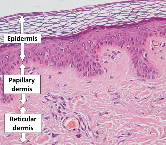
The dermis is not a uniform layer; it's divided into two distinct regions: a superficial papillary layer and a deep reticular layer. Let's examine each one in detail.
The Papillary Layer: Anchoring and Nourishing
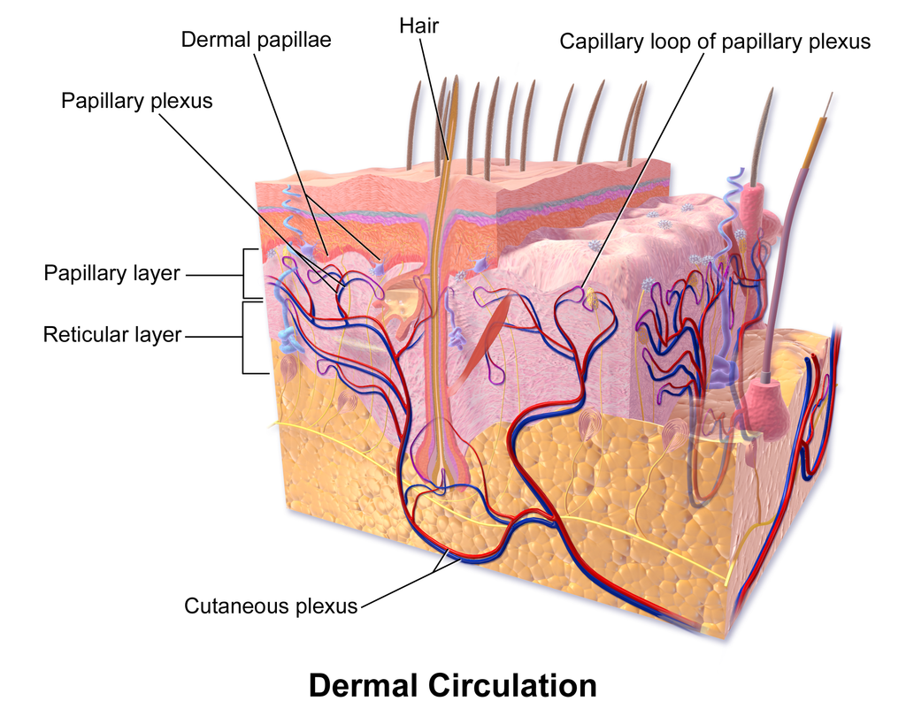
The papillary layer is the upper region of the dermis, making up about 20% of its total thickness. It's composed of loose, areolar connective tissue. Its most distinctive feature is the presence of dermal papillae, which are finger-like projections that extend upward and interlock with the ridges of the epidermis. This intricate connection firmly anchors the two layers together, preventing them from sliding past one another. In the thick skin of your hands and feet, these papillae form the epidermal ridges you know as fingerprints.
This layer is crucial for the survival of the epidermis. Let's explore its contents and functions.
Integumentary System Lecture CHAPTER 5
The "Integumentary System Lecture" from nursing2nurture offers a clear, student-focused explanation of the papillary layer.
Please watch the segment covering the papillary layer. Pay close attention to the description of the dermal papillae, the consequence of disrupting the collagen fibers (blisters), and the types of structures housed within this layer.
As the video explained, the key functions and structures within the papillary layer are:
- Capillaries: The dermal papillae contain an extensive network of capillaries known as the papillary plexus. Since the epidermis is avascular, this is the blood supply from which oxygen and nutrients diffuse to nourish the cells of the stratum basale.
- Sensory Receptors: This layer contains nerve endings and specialized touch receptors called Meissner's corpuscles (or tactile corpuscles). These are responsible for detecting light touch, which is why areas like your fingertips and lips are so sensitive.
- Defensive Cells: Phagocytes, a type of immune cell, are also present, ready to attack any bacteria or pathogens that manage to breach the epidermis.
To solidify this, let's review a more detailed textual description.
Layers of the Skin | Anatomy and Physiology I
The Lumen Learning article provides a concise written summary of the papillary layer's components.
Please read the section titled "Papillary Layer." This will reinforce what you learned from the video about the tissue type, the cells it contains (fibroblasts, phagocytes), and its sensory structures.
The Reticular Layer: Strength and Elasticity
Beneath the papillary layer lies the much thicker reticular layer, which accounts for about 80% of the dermis. This layer is made of dense irregular connective tissue, which contains a meshwork of collagen and elastic fibers.
- Collagen fibers give the skin incredible tensile strength, preventing it from being torn by pulling forces. They are also hydrophilic, meaning they attract and bind water, which helps keep the skin hydrated and firm. This property is what you are assessing when you check for skin turgor.
- Elastic fibers allow the skin to stretch and recoil back to its original shape. If the skin is stretched beyond its elastic limits, such as during rapid weight gain or pregnancy, these fibers can tear, resulting in stretch marks (striae).
This layer is where most of the skin's accessory structures are anchored.
Integumentary System Lecture CHAPTER 5
Let's return to the nursing2nurture lecture to understand the reticular layer.
Watch the next segment of the video, which describes the reticular layer. Note the difference in fiber content compared to the papillary layer, the explanation of stretch marks, and the mention of the deeper sensory receptors found here.
The reticular layer is the functional heart of the dermis. It contains:
- Blood Vessels: A larger network of blood vessels called the cutaneous plexus is located at the border between the reticular layer and the underlying hypodermis. Branches from this plexus supply the glands, follicles, and other structures in the dermis.
- Glands and Follicles: Sweat glands, sebaceous (oil) glands, and hair follicles all originate in this layer. We'll explore these in our next lessons.
- Nerve Fibers: This layer has a rich nerve supply, including deep pressure receptors called Pacinian corpuscles (or lamellated corpuscles). Their layered, onion-like structure makes them sensitive to pressure and vibration.
To round out your understanding, let's turn to a final reading.
Anatomy Lecture Notes Section 1: The Integumentary System
These lecture notes from San Diego Miramar College provide an excellent, detailed breakdown of the reticular layer's contents.
Read the section titled "The Reticular Layer". Focus on the list of elements it contains, which provides a comprehensive summary of its complex structure.
Conclusion
In this lesson, we've explored the two layers of the dermis, the supportive core of our skin. You've learned how its structure directly relates to its critical functions in nourishment, sensation, and physical resilience.
Here are the key takeaways:
- The dermis is divided into the superficial papillary layer and the deep reticular layer.
- The papillary layer is composed of loose, areolar connective tissue. Its dermal papillae lock it into the epidermis, and its capillary plexus nourishes the epidermal cells. It also contains Meissner's corpuscles for sensing light touch.
- The reticular layer is composed of dense irregular connective tissue, providing strength (collagen fibers) and elasticity (elastic fibers). It houses the skin's accessory structures like glands and hair follicles, as well as Pacinian corpuscles for sensing deep pressure.
- The health of the collagen and elastic fibers, along with their ability to hold water, is directly related to skin turgor and the formation of wrinkles and stretch marks.
In our next lesson, we will focus on the accessory structures of the skin, such as hair, hair follicles, and nails, which are anchored deep within the reticular layer we just discussed.