Hello! Welcome to your first lesson in Anatomy and Physiology. Given your tight schedule for the upcoming exam, we'll dive straight into the first topic of the Integumentary System.
This lesson focuses on the outermost layer of your skin, the epidermis. Our goal is to identify its distinct layers and describe the major types of cells you'd find in each one. Understanding this structure is fundamental for your nursing studies, as it's the body's first line of defense and is central to topics like wound healing, drug administration, and assessing skin conditions—all of which you'll encounter in a clinical setting.
The Epidermis: An Overview
The epidermis is the most superficial layer of the skin. It is a shield, protecting the deeper tissues from water loss, mechanical injury, and pathogens. Structurally, it is classified as keratinized stratified squamous epithelium. That's a mouthful, but it simply means it's made of multiple layers (stratified) of flattened cells (squamous) that are hardened with a protein called keratin.
A crucial feature of the epidermis is that it is avascular, meaning it has no blood vessels. Its cells get oxygen and nutrients entirely by diffusion from the capillaries in the dermis below.
The vast majority of cells in the epidermis are keratinocytes. As their name suggests, they produce keratin, a tough, fibrous protein that gives the skin its protective, water-resistant properties. Keratinocytes are constantly produced in the deepest layer of the epidermis and are gradually pushed toward the surface, maturing and dying along the way. This journey from birth to being sloughed off the surface takes about 4-6 weeks.
To begin, let's get a comprehensive overview of the layers and the keratinocyte life cycle.
Layers of the Skin | Anatomy and Physiology I
This reading from Lumen Learning provides a concise introduction to the epidermis and the journey of a keratinocyte.
Please read the first few paragraphs, from the beginning down to the end of the paragraph about keratinocytes. This will set the stage for our detailed tour of the epidermal layers.
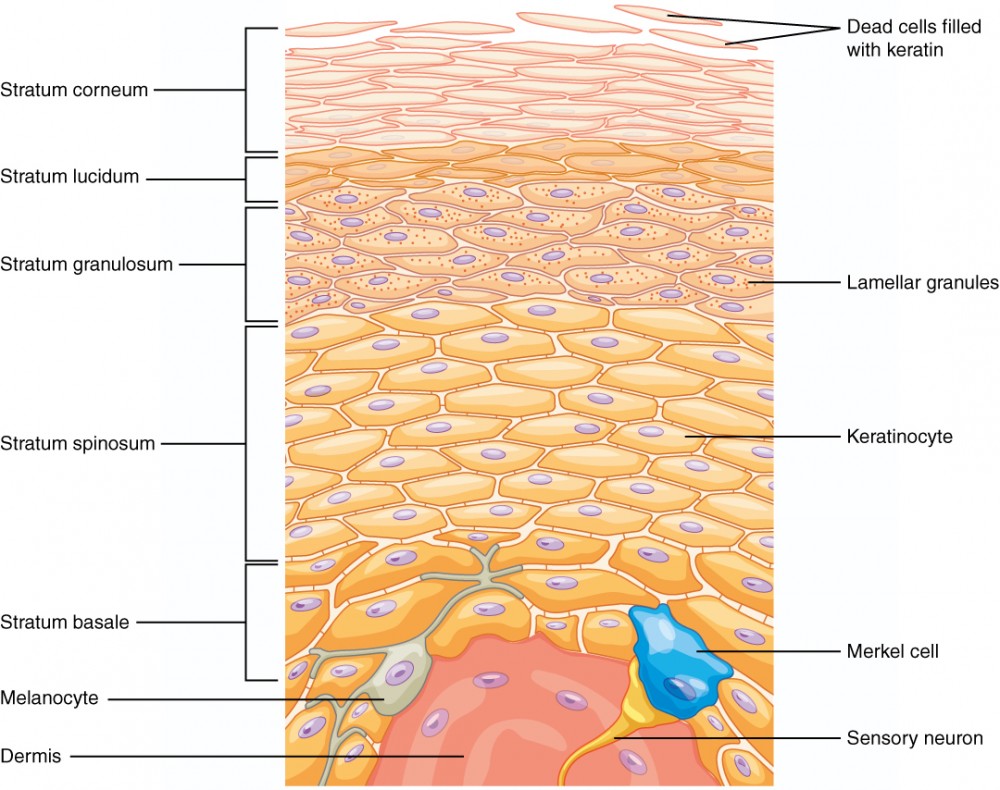
Now that you have a general idea, let's look at a diagram that we will refer to throughout the lesson. It illustrates the different layers, or strata, of the epidermis.
A Quick Tour of the Epidermal Layers
To get a quick, dynamic overview of these layers and the cells within them, let's watch a short video. It also provides a helpful mnemonic to remember the layers in order from superficial to deep.
This video from Amoeba Sisters provides a rapid tour of the epidermis, introducing keratinocytes, the five strata, and the other major cell types.
Watch the segment from the introduction of keratinocytes to the end of the epidermis section. Pay close attention to the mnemonic "Can Lemurs Get Some Bamboo?" which corresponds to the layers from top to bottom: Corneum, Lucidum, Granulosum, Spinosum, Basale.
The Strata of the Epidermis: From Deep to Superficial
The video gave a great top-down tour. Now, we will examine the layers more closely, following the path a keratinocyte takes as it matures. We'll start from the bottom (deepest layer) and work our way up to the top (most superficial layer).
We'll use the Lumen Learning article again for a more detailed look at each stratum.
1. Stratum Basale (or Stratum Germinativum)
This is the deepest layer, attached to the underlying dermis. It consists of a single layer of cuboidal-shaped basal cells, which are stem cells. This is the mitotic layer, where new keratinocytes are constantly being "born" (hence the alternative name, germinativum). The rate of cell division here is crucial for skin regeneration and wound healing. In aging individuals, a slower mitotic rate in this layer contributes to thinner skin and slower healing, a common concern in geriatric care.
Two other important cell types reside here:
- Melanocytes: These cells produce the pigment melanin, which is responsible for skin color. Melanin is transferred to the newly formed keratinocytes, where it clusters around the nucleus to protect the cell's DNA from UV radiation damage.
- Merkel cells: These are sensory receptors for touch, connecting to nerve endings in the dermis.
Layers of the Skin | Anatomy and Physiology I
Let's zoom in on this foundational layer.
In the Lumen Learning article, find the section titled "Stratum Basale" and read from that heading down to the end of the paragraph describing melanocytes.
2. Stratum Spinosum
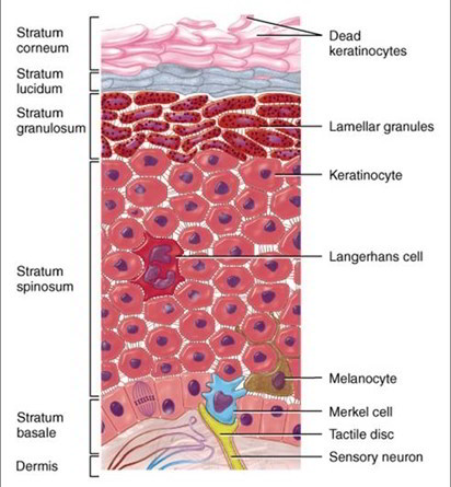
As new cells form in the stratum basale, they push older ones up into the stratum spinosum, which is typically 8-10 cell layers thick. The cells here begin to flatten and produce keratin. They are interconnected by cellular junctions called desmosomes, which give the cells a "spiny" appearance on microscope slides (this is an artifact of the staining process).
This layer also contains scattered Langerhans cells (a type of dendritic cell). These are immune cells that act as macrophages, engulfing bacteria and foreign particles to protect the body.
Layers of the Skin | Anatomy and Physiology I
Now, let's look at the "spiny" layer.
Read the section titled "Stratum Spinosum," from that heading to the end of the section.
3. Stratum Granulosum
This is the "grainy layer," consisting of 3-5 layers of flattened keratinocytes. By this point, the cells have stopped dividing and are producing large amounts of keratin. They also produce granules containing a lipid-rich secretion that acts as a water sealant. As the cells fill with keratin, their nuclei and organelles begin to disintegrate, and the cells die. This marks the transition between the deeper, living layers and the superficial, dead layers.
Layers of the Skin | Anatomy and Physiology I
Next is the grainy, transitional layer.
Please read the short section on the "Stratum Granulosum," from its heading to the end of the section.
4. Stratum Lucidum
This "clear layer" is only found in thick skin, such as the palms of your hands and the soles of your feet. It consists of a few rows of clear, flat, dead keratinocytes.
5. Stratum Corneum
Finally, we reach the most superficial layer, the stratum corneum. It consists of 15-30 layers of dead, flattened, fully keratinized cells. These durable, water-resistant cells form a tough barrier against abrasion, pathogens, and dehydration. Cells from this layer are continuously shed and replaced by cells pushed up from below.
Layers of the Skin | Anatomy and Physiology I
We'll finish our tour with the final two layers.
Read the last two sections on the epidermal layers: "Stratum Lucidum" and "Stratum Corneum."
A Closer Look at the Cells
We've discussed the four main cell types as they appear in the layers, but let's watch one more short video to consolidate your understanding of their specific roles.
The video "Cells of the Epidermis" by A&P Made Easy clearly explains the function of each cell type.
Watch the following segments to review the four key cells: Keratinocytes: the primary structural cells. Dendritic (Langerhans) cells: the immune guardians. Tactile (Merkel) cells: the touch sensors. Melanocytes: the pigment producers.
Conclusion
In this lesson, we have dissected the epidermis, the protective outer shield of our body. We've seen how it is a dynamic, multi-layered structure constantly regenerating itself.
Here are the key takeaways for your exam preparation:
- The epidermis has five layers in thick skin and four in thin skin. From deep to superficial, they are: Stratum Basale, Stratum Spinosum, Stratum Granulosum, Stratum Lucidum (thick skin only), and Stratum Corneum.
- The life of a keratinocyte is a journey of maturation from the basal layer to the surface, ending as a dead, protective cell in the stratum corneum.
- The epidermis houses four major cell types with distinct functions:
- Keratinocytes: Provide structure and water resistance.
- Melanocytes: Produce melanin for UV protection.
- Langerhans cells: Provide immune defense.
- Merkel cells: Function in touch sensation.
In our next lesson, we will move deeper into the skin to explore the dermis. This is the layer that provides the epidermis with nutrients and contains many of the skin's accessory structures, like glands and hair follicles.