Hello! In our last lesson, we explored the dermis and its two layers, the papillary and reticular layers, which form the supportive core of the skin. Now, we will build directly on that knowledge by examining the accessory structures that are anchored deep within the dermis: hair, hair follicles, and nails.
This lesson is designed to help you describe the structure and functions of these important epidermal derivatives. For your nursing career, understanding these structures is not just an anatomical exercise. The condition of a patient's hair and nails can provide valuable clues about their nutritional status, oxygenation, and overall systemic health—insights you will find useful, perhaps even in your current work at the retirement home.
Hair and Hair Follicles: More Than Just Decoration
Hair is a filament composed of dead, keratinized cells that grows from an organ called a hair follicle. These follicles are essentially tubes formed by an invagination, or folding-in, of the epidermis that extends down into the dermis.
Let's begin by watching a video that provides a clear overview of the anatomy of both hair and hair follicles.
Hair, Hair follicle, and Nail Structure
This video from "A&P Made Easy" will introduce you to hair as a derivative of the epidermis and then walk you through the key anatomical structures of a hair follicle.
First, watch the brief introduction from the beginning to understand how follicles are formed. Then, continue with the detailed breakdown of the hair follicle's structure from hair follicle anatomy. As you watch, pay close attention to the terms for the different parts of the follicle and the layers of the hair itself.
As the video explained, the anatomy of hair and its follicle is quite intricate. Let's break down the key components, using a diagram to visualize them.
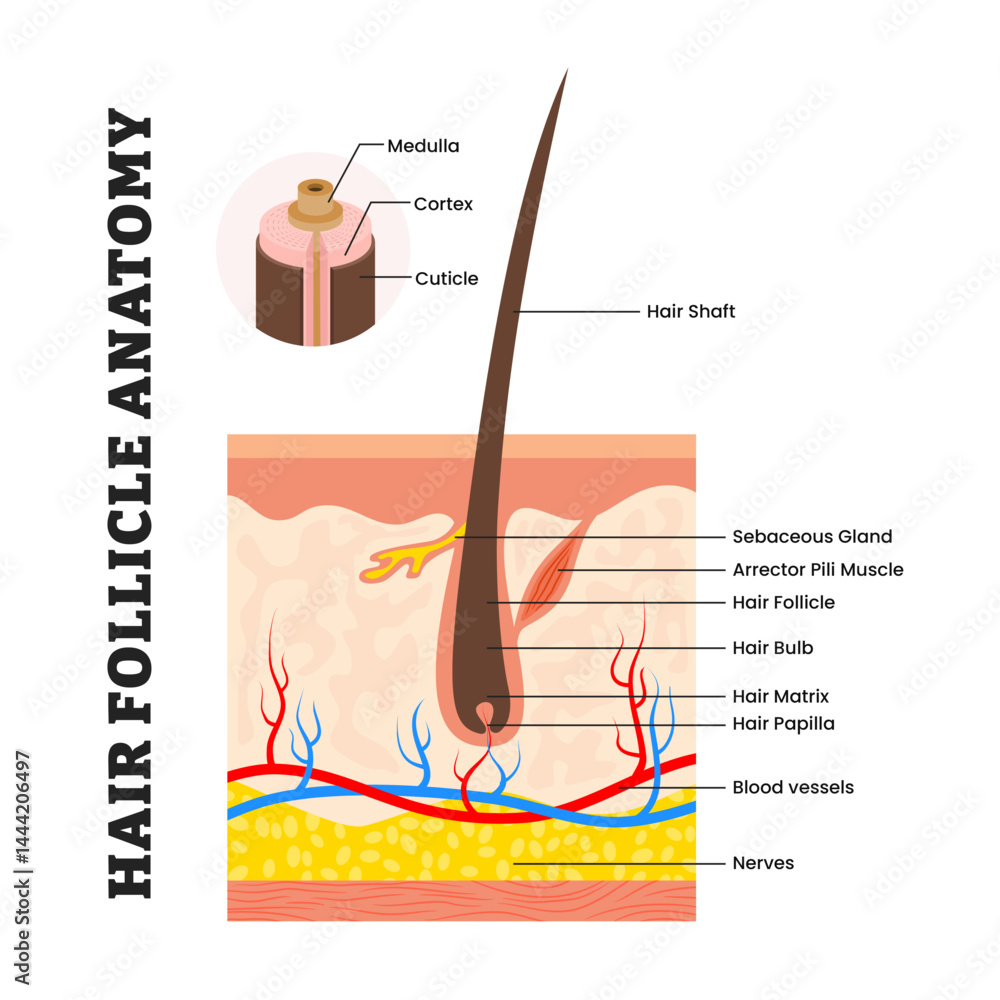
Based on the video and the diagram above, here are the essential structures to know:
- Hair Follicle: The sheath of epidermal and dermal tissue that contains the hair. Its base is expanded into a hair bulb.
- Hair Papilla: A small cone of dermal tissue that protrudes into the hair bulb. It contains capillaries that supply nutrients to the growing hair.
- Hair Matrix: A layer of actively dividing epithelial cells located in the hair bulb, surrounding the papilla. This is the growth center of the hair. As new cells are produced, older ones are pushed upward, die, and become keratinized to form the hair itself.
- Hair Root and Shaft: The root is the portion of the hair enclosed within the follicle, while the shaft is the part that projects from the surface of the skin. The shaft is what we see, and it is biologically dead.
- Layers of the Hair: The hair itself has three concentric layers:
- Medulla: The central core.
- Cortex: The bulky middle layer, which contains the pigments (melanin) that give hair its color.
- Cuticle: The outermost layer, formed from a single layer of overlapping cells that look like shingles on a roof. It provides strength and protection.
Associated Structures and Functions
The hair follicle doesn't exist in isolation. It's connected to several other important structures that contribute to the skin's overall function.
Dr. John Campbell's video on hair provides excellent explanations for the structures that support the follicle.
Watch the following three short segments: Dermal papilla: This explains the crucial role of the blood supply in nourishing the growing hair. Hair erector muscle: This covers the function of the arrector pili muscle. Hair root plexus: This explains the sensory function associated with hair.
Let's summarize these key associated structures and the primary functions of hair:
- Arrector Pili Muscle: A tiny band of smooth muscle attached to the follicle. When it contracts (in response to cold or fear), it pulls the hair follicle upright, causing "goosebumps." This traps a layer of air for insulation, though its effect is minimal in humans compared to furred animals.
- Hair Root Plexus: A network of sensory nerve endings that wraps around the base of each follicle. This allows you to feel even a light touch that moves the hair, acting as a sensitive receptor.
- Sebaceous Gland: This oil gland almost always opens into a hair follicle. It secretes an oily substance called sebum that lubricates the hair and skin. We will cover this gland in detail in our next lesson.
The primary functions of hair include:
- Protection: Hair on the scalp guards against UV radiation and cushions against minor impacts.
- Sensation: The hair root plexus detects movement of the hair shaft.
- Insulation: Helps to retain heat, especially on the scalp.
Nails: Protective Plates
Nails are the second accessory structure we will cover. Like hair, they are a modification of the epidermis and are composed of hard, keratinized cells.

Let's watch a final video segment to walk through the anatomy of the nail.
Hair, Hair follicle, and Nail Structure
We return to the "A&P Made Easy" video for a concise explanation of nail structure.
Please watch the section covering nail anatomy. This will introduce you to the key terminology for the parts of a nail.
Here is a breakdown of the main parts of the nail:
- Nail Plate (or Body): The visible, attached portion of the nail.
- Free Edge: The portion of the nail that grows out away from the body.
- Nail Root: The proximal part of the nail embedded in the skin.
- Nail Bed: The layer of epidermis on which the nail plate rests.
- Nail Matrix: The thickened, proximal part of the nail bed responsible for nail growth. The cells here are actively dividing, and as they become keratinized, they are pushed distally, causing the nail to grow.
- Lunula: The white, crescent-shaped region at the base of the nail. It is the visible part of the nail matrix.
- Eponychium: Commonly known as the cuticle, this is the fold of stratum corneum at the proximal end of the nail.
Clinical Significance of Nails
Nails serve to protect the sensitive tips of our fingers and toes and can act as "tools" to pick up small objects. However, for a healthcare professional, their most important function is as an indicator of health.
The article from Bio LibreTexts has an excellent section on the connection between nails and health, which is highly relevant to your nursing studies.
Please read the section titled Nails and Health. Pay close attention to the description of the blanch test (capillary refill test) and what changes in nail color might signify.
As you just read, observing the nails is a quick, non-invasive way to assess a patient. A bluish or purple nail bed can indicate poor oxygenation (cyanosis), while the capillary refill test (blanch test) helps you assess blood flow to the extremities. Changes in nail shape, thickness, or texture can also signal nutritional deficiencies or systemic diseases.
Conclusion
In this lesson, we have examined the accessory structures of the skin: hair, hair follicles, and nails. You've learned how these structures are formed from the epidermis and anchored in the dermis, and you've seen how their condition can offer important diagnostic clues.
Here are the main takeaways:
- Hair is a filament of dead, keratinized cells produced by the hair follicle. Growth occurs at the hair matrix within the hair bulb, which is nourished by the hair papilla.
- Associated structures like the arrector pili muscle (goosebumps) and the hair root plexus (sensation) add to the skin's functionality.
- Nails are hard plates of keratinized cells that protect the ends of the digits. They grow from the nail matrix at the base of the nail.
- Both hair and nails are clinically significant. Their appearance can reflect a person's oxygenation, nutritional status, and overall health.
In our next lesson, we will complete our look at the skin's accessory structures by focusing on the glands of the skin: sebaceous (oil) glands and sudoriferous (sweat) glands.