Welcome to our next lesson. In our previous session, we examined hair, follicles, and nails, and you learned that sebaceous glands are closely associated with hair follicles. We will now expand on that by exploring all the major glands of the skin.
This lesson focuses on comparing the structure, function, and location of the skin's two main types of glands: sebaceous (oil) glands and sudoriferous (sweat) glands. A solid understanding of these glands is essential for a nursing career, as they are fundamental to thermoregulation, skin integrity, and protection from infection. Observing changes in their function can provide clues about a patient's health, particularly concerning hormonal shifts or dehydration.
Glands of the Skin: An Overview
The skin contains several types of exocrine glands, which secrete substances onto an epithelial surface. They all originate from the epidermis but extend down into the dermis. The two primary categories are sweat glands and oil glands.
Let's begin with a comprehensive video from RegisteredNurseRN that covers the key characteristics of all these glands. This will provide a strong foundation for the rest of the lesson.
Skin Glands Anatomy: Sweat Glands, Sebaceous Glands Integumentary System
This video offers a complete overview of the skin's glands, from the different types of sweat glands to the oil-producing sebaceous glands and their unique secretion methods.
Please watch the entire video (full video). As you watch, focus on distinguishing between the two main types of sweat glands (eccrine and apocrine) and note the key differences between sweat glands and sebaceous glands in terms of their secretions and how they release them.
Now that you have a general overview, let's break down the details for each type of gland.
Sudoriferous (Sweat) Glands
As the name implies, these glands produce sweat. They are also called sudoriferous glands and are crucial for regulating body temperature. There are two distinct types: eccrine and apocrine glands.

1. Eccrine Sweat Glands
These are the most numerous sweat glands and are the key players in thermoregulation.
- Structure and Location: Eccrine glands are simple, coiled tubular glands located in the dermis. They are found all over the body but are most concentrated on the palms, soles of the feet, and forehead. Their ducts open directly onto the skin's surface through a funnel-shaped opening called a pore.
- Function and Secretion: They secrete a watery, hypotonic fluid we call sweat. This sweat is about 99% water, with some salts (like sodium chloride), metabolic wastes, and an antimicrobial peptide called dermcidin. Their primary function is evaporative cooling. When body temperature rises, these glands release sweat. As the sweat evaporates from your skin, it takes heat with it, cooling the body down. This process relies on the principle of the "heat of vaporization," a concept you may remember from your chemistry course.
To better understand this cooling mechanism, please read the following short text.
3.13: Sweat and Sebaceous Glands
This section from Med LibreTexts provides an excellent explanation of how eccrine glands facilitate evaporative cooling.
Read the section titled Eccrine Glands. Focus on how the evaporation of sweat removes heat from the body.
2. Apocrine Sweat Glands
Apocrine glands function differently from eccrine glands and are associated with hair follicles.
- Structure and Location: These glands are larger than eccrine glands and are found in the axillary (armpit) and anogenital regions. Unlike eccrine glands, their ducts empty into hair follicles.
- Function and Secretion: Apocrine glands do not become fully functional until puberty. They secrete a thicker, milky or yellowish substance that contains the same components as eccrine sweat, plus fatty substances and proteins. The secretion itself is odorless, but when bacteria on the skin decompose its organic molecules, it produces body odor. Apocrine glands are activated by nerve fibers during times of pain, stress, and sexual arousal, but they play a minimal role in thermoregulation.
The video you watched also mentioned modified apocrine glands, which include:
- Ceruminous glands: Found in the lining of the external ear canal; their secretion mixes with sebum to produce cerumen, or earwax.
- Mammary glands: Specialized glands that secrete milk.
Secretion Method: Merocrine
Both eccrine and apocrine sweat glands are classified as merocrine glands. This means they release their products via exocytosis, a process where vesicles fuse with the cell membrane to release their contents. The cell itself remains completely intact during this process.
Sebaceous (Oil) Glands
Next, we turn to the glands that were briefly mentioned in our last lesson: the sebaceous glands.
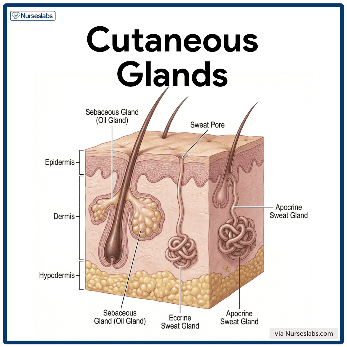
- Structure and Location: Sebaceous glands are simple branched alveolar glands. They are found all over the body except on the palms and soles. They typically empty their secretions into a hair follicle, although some open directly onto the skin surface in hairless areas.
- Function and Secretion: These glands produce an oily substance called sebum. Sebum is a mixture of lipids and cell fragments. It serves several functions:
- Lubrication: It softens and lubricates the hair and skin, preventing them from becoming dry and brittle.
- Waterproofing: It helps slow water loss from the skin.
- Antibacterial: It has bactericidal (bacteria-killing) properties.
Like apocrine glands, sebaceous glands are activated at puberty in response to hormones, which is why skin tends to become oilier during adolescence.
Secretion Method: Holocrine
Sebaceous glands have a different method of secretion. They are classified as holocrine glands. In this process, the secretory cells accumulate their product (sebum) until they become so engorged that they rupture and die. The entire dead cell, filled with its lipid product, becomes the secretion itself. This is a key distinction from the merocrine secretion of sweat glands.
Comparison Summary
To consolidate your understanding and prepare for your exam, here is a table comparing the key features of these glands.
| Feature | Eccrine Sweat Gland | Apocrine Sweat Gland | Sebaceous (Oil) Gland |
|---|---|---|---|
| Product | Watery sweat (water, salts, waste) | Viscous sweat (water, lipids, protein) | Sebum (lipids, cell fragments) |
| Function | Thermoregulation (cooling) | Scent/pheromonal signaling | Lubrication, waterproofing, antibacterial |
| Location | Most of the body; dense on palms/soles | Axillary and anogenital regions | Most of the body; absent on palms/soles |
| Duct Opening | Directly onto skin surface (pore) | Into a hair follicle | Usually into a hair follicle |
| Secretion Method | Merocrine (exocytosis) | Merocrine (exocytosis) | Holocrine (cell ruptures) |
| Activation | Soon after birth | Puberty | Puberty |
Conclusion
In this lesson, we have differentiated the glands of the skin. You now know that the integumentary system contains sudoriferous (sweat) glands for cooling and communication, and sebaceous (oil) glands for lubrication and protection.
Here are the key takeaways:
- Eccrine glands are the body's primary cooling mechanism, releasing watery sweat directly onto the skin.
- Apocrine glands, activated at puberty, release a thicker sweat into hair follicles, which is linked to body odor.
- Sebaceous glands produce oily sebum via a destructive holocrine process, waterproofing and lubricating the skin and hair.
- Sweat glands use a non-destructive merocrine secretion method, where the cell remains intact.
In our next lesson, we will examine how the entire integumentary system, including these glands, responds to injury and the effects of aging. Understanding how these structures function in a healthy state is crucial for recognizing what goes wrong during these processes.