Welcome back! In our last lesson, we explored the anatomy of the femur and patella, concluding with their articulation at the knee joint. We noted how the large condyles of the femur are shaped to meet the bones of the lower leg.
Today, we will complete our examination of the leg's bony framework by focusing on those two bones: the tibia and the fibula. Our goal is to identify the tibia and fibula and their key bony landmarks. You will learn to distinguish between these two bones, understand their respective roles in weight-bearing and stability, and identify the specific features that are crucial for muscle attachment and joint formation at both the knee and the ankle. This knowledge is fundamental for assessing common lower limb injuries you will encounter in your nursing career, from shin splints to ankle fractures.
The Bones of the Lower Leg: An Overview
The lower leg, the region between the knee and the ankle, contains two bones: the tibia and the fibula.
- The tibia is the larger, medial bone. It is the main weight-bearing bone of the lower leg. You can easily feel its anterior surface just under the skin of your shin.
- The fibula is the thinner, lateral bone. It does not bear significant weight but is a critical site for muscle attachment and plays a key role in stabilizing the ankle joint.
A common mnemonic to remember their relationship is: "Never tell a Little fib." The "fib" reminds you of the fibula, "little" reminds you it's the smaller bone, and the "L" in "little" helps you remember it is always on the lateral side of the leg.
Let's begin our detailed examination with the tibia.
The Tibia (Shin Bone)
The tibia is the second longest bone in the body, after the femur. Its size reflects its crucial function: transmitting the body's weight from the femur to the foot. We will identify its landmarks by working from its proximal end at the knee down to its distal end at the ankle.
This video will serve as our primary guide for identifying the tibia's features.
Tibia and Fibula Anatomy of Leg Bones | Anatomy & Physiology
The video "Tibia and Fibula Anatomy" by RegisteredNurseRN provides a very clear, systematic walkthrough of the tibial landmarks.
Please watch the segment from the tibia section. As you watch, focus on locating and understanding the function of the following structures: Proximal End: Medial and Lateral Condyles, Articular Surfaces, Intercondylar Eminence Shaft: Tibial Tuberosity, Anterior Crest Distal End: Medial Malleolus, Fibular Notch, Articular Surface
To summarize the key landmarks of the tibia:
- Proximal Tibia (at the knee):
- Medial and Lateral Condyles: Two large, flattened platforms on the superior end of the tibia. Their smooth top surfaces, the articular surfaces, articulate with the medial and lateral condyles of the femur.
- Intercondylar Eminence: A raised area between the two condyles that fits into the intercondylar fossa of the femur. It provides stability to the knee joint by serving as an attachment point for the cruciate ligaments.
- Shaft of the Tibia:
- Tibial Tuberosity: A prominent rough bump on the anterior surface, just below the condyles. This is the attachment site for the patellar ligament.
- Anterior Crest: The sharp ridge that runs down the anterior surface of the tibia. This is the part of the bone commonly known as the "shin."
- Distal Tibia (at the ankle):
- Medial Malleolus: A bony prominence on the medial side of the ankle. This is the bump you can feel on the inner side of your ankle.
- Fibular Notch: A shallow depression on the lateral side of the distal tibia, which articulates with the fibula.
The Fibula
Now let's turn our attention to the fibula. While it is much thinner than the tibia and isn't directly involved in bearing weight, it is essential for muscle attachment and forming the stable outer wall of the ankle joint.
For the fibula, we'll use the video "Tibia and Fibula" from The Noted Anatomist. This video offers another excellent perspective and includes a clinically relevant point about the fibular neck.
First, watch the segment introducing the fibula and its proximal landmarks from this part. Note the head and neck of the fibula and the important nerve that passes near the neck. Next, watch the segment on the distal fibula from here. This will identify the lateral malleolus.
The key landmarks of the fibula are simpler:
- Head: The enlarged proximal end, which articulates with the lateral condyle of the tibia.
- Neck: The constricted area just distal to the head. As the video mentions, the common peroneal (or fibular) nerve wraps around the fibular neck, making it vulnerable to injury if the neck is fractured. Damage to this nerve can impair the ability to lift the foot (a condition known as "foot drop").
- Lateral Malleolus: The pointed distal end of the fibula. It forms the bony bump on the outer side of your ankle and is larger and more prominent than the medial malleolus of the tibia.
Connecting the Tibia and Fibula
The tibia and fibula do not function in isolation. They are held together by a strong membrane and articulate at both their proximal and distal ends.
Tibia and Fibula Anatomy with Labeled Diagram
The article "Tibia and Fibula Anatomy" from SimpleNursing provides a concise explanation of the membrane that unites these two bones.
Please read the short section titled Interosseous Membrane.
As the reading explains, the shafts of the tibia and fibula are connected by the interosseous membrane, a dense sheet of connective tissue. This membrane not only binds the bones together but also serves as an attachment surface for several muscles of the leg.
The image below shows the landmarks of both bones in anterior and posterior views, including the interosseous membrane connecting them.
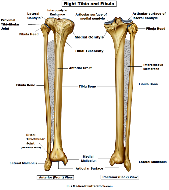
The final, and perhaps most critical, relationship between these bones is at the ankle. The medial malleolus of the tibia and the lateral malleolus of the fibula form a deep socket, or mortise, that cups the talus (one of the ankle bones). This mortise joint is what allows you to dorsiflex and plantarflex your foot.
Practice Identification
To consolidate your knowledge, use the unlabeled diagram below to test yourself. Try to identify as many landmarks as you can on both the tibia and fibula. You can then refer back to the labeled diagram above to check your answers. This is an excellent way to prepare for your exam.

Conclusion
In this lesson, we have successfully identified the bony landmarks of the tibia and fibula. We've established their respective roles, with the tibia serving as the primary weight-bearing bone and the fibula providing stability and muscle attachment points. By tracing their features from the knee to the ankle, we have completed our anatomical survey of the bones of the leg.
Key Takeaways:
- The tibia is the large, medial bone of the lower leg. Its key landmarks include the medial and lateral condyles, tibial tuberosity, anterior crest, and medial malleolus.
- The fibula is the slender, lateral bone. Its main features are the head, neck, and lateral malleolus.
- The two bones are connected along their shafts by the interosseous membrane.
- Distally, the medial malleolus (tibia) and lateral malleolus (fibula) form the ankle mortise, which articulates with the talus bone of the foot.
Having now arrived at the ankle joint, our next lesson will take the final step in our journey through the lower limb. We will identify the bones of the ankle and foot: the tarsals, metatarsals, and phalanges. We will begin with the talus, the bone that fits snugly into the mortise we just described.