Welcome to our next session focusing on the appendicular skeleton. In the previous lesson, we examined the pelvic girdle and learned to distinguish between the male and female pelvis. We concluded by noting that the hip joint is formed by the articulation of the os coxa's acetabulum with the head of the femur.
Today, we will continue our journey down the lower limb to fulfill our next learning outcome: to identify the femur and patella and their key bony landmarks. We will explore the femur, the single bone of the thigh and the longest, heaviest, and strongest bone in the human body. Then, we will examine the patella, or kneecap, a unique bone that plays a crucial role in the function of the knee joint. For your future work in nursing, a solid understanding of these bones is essential, as they are frequently involved in common conditions you'll encounter, from hip fractures in elderly patients to knee injuries in athletes.
Let's begin with the femur.
The Femur: Bone of the Thigh
The femur is remarkable not just for its size but for its critical role in supporting the body's weight and enabling locomotion. It extends from the hip to the knee. We will examine its structure in three parts: the proximal end (articulating at the hip), the shaft, and the distal end (articulating at the knee).
This image provides an excellent overview of the femur in both anterior and posterior views, showing its relationship with the hip bone and the bones of the lower leg.

To systematically identify the femur's many landmarks, we will use a detailed video guide.
The video "Femur" by The Noted Anatomist provides a clear, step-by-step tour of the bone's features. We will start with the proximal end.
Please watch the segment from the proximal femur. As you watch, pay close attention to the location and description of the following landmarks: head, fovea capitis, neck, greater and lesser trochanters, intertrochanteric line, and intertrochanteric crest.
To recap, the key landmarks of the proximal femur are:
- Head: The smooth, rounded top that fits into the acetabulum of the hip bone.
- Fovea Capitis: A small pit on the head for the attachment of a ligament (ligamentum teres).
- Neck: The constricted region just below the head that connects it to the shaft.
- Greater and Lesser Trochanters: Large and small bony projections, respectively, that serve as important attachment sites for muscles of the hip and thigh. The greater trochanter is lateral, and the lesser trochanter is posteromedial.
- Intertrochanteric Line (anterior) and Intertrochanteric Crest (posterior): Ridges that run between the two trochanters, marking the junction of the neck and shaft.
Clinical Relevance: Femoral Neck Fractures
Given your work in a retirement home, you are likely aware that "broken hips" are a common and serious injury among the elderly. These are most often fractures of the femoral neck. Due to changes in bone density with age (osteoporosis), a fall that might only cause a bruise in a younger person can result in a devastating fracture in an older adult.
Anatomy, Bone Markings - StatPearls - NCBI Bookshelf - NIH
This excerpt from a StatPearls article on bone markings provides a concise clinical overview of femoral neck fractures.
Please read the brief section titled Femoral Neck Fracture. This will connect the anatomy you're learning to a real-world clinical scenario.
The Femoral Shaft and Distal End
Now, let's move down the bone to its shaft and distal end.
Continuing with the same video, we'll now look at the features of the shaft and the distal end, which forms the knee joint.
First, watch the segment on the shaft's posterior features from this clip. This covers the gluteal tuberosity and the prominent linea aspera. Next, watch the portion covering the distal end from here. This section identifies the medial and lateral condyles, the intercondylar notch (or fossa), the medial and lateral epicondyles, the adductor tubercle, and the patellar surface.
Let's summarize these features:
- Shaft: The long, central part of the bone. Its most notable feature is the linea aspera ("rough line"), a prominent ridge on the posterior surface that serves as an attachment point for many muscles. Proximally, it is continuous with the gluteal tuberosity, where the gluteus maximus muscle attaches.
- Distal End: This end is wider to help distribute weight at the knee joint.
- Medial and Lateral Condyles: Smooth, rounded surfaces that articulate with the tibia of the lower leg.
- Medial and Lateral Epicondyles: Roughened projections on the outer surfaces of the condyles that serve as attachment points for ligaments of the knee.
- Adductor Tubercle: A small bump on the superior aspect of the medial epicondyle for the adductor magnus muscle.
- Intercondylar Fossa: A deep depression on the posterior side, between the condyles.
- Patellar Surface: A smooth, shallow depression on the anterior side, where the patella articulates.
The Patella: The Kneecap
Having identified the patellar surface on the femur, we can now turn our attention to the patella itself. The patella is the largest sesamoid bone in the body, meaning it is a bone that forms within a tendon. It is embedded in the tendon of the quadriceps femoris muscle group. Its primary functions are to protect the anterior aspect of the knee joint and to increase the leverage of the quadriceps muscle.
Patella Anatomy | Patella Kneecap Bone Review
The video "Patella Anatomy" by RegisteredNurseRN provides an excellent guide to this small but important bone.
First, watch the introduction from the beginning to understand the patella's classification and function. Next, watch the segment detailing the anterior surface from this part. This will identify the base, apex, and medial/lateral borders. Finally, watch the description of the posterior surface from the posterior view. Focus on the smooth articular facets that glide on the condyles of the femur. Notice the tip for telling a left from a right patella based on the size of the facets.
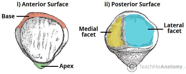
Here are the key landmarks of the patella, which you can also see in the image below:
- Anterior Surface: Is rough and convex.
- Base: The broad, superior border where the quadriceps tendon attaches.
- Apex: The pointed, inferior tip that connects to the patellar ligament (which then attaches to the tibia).
- Posterior Surface: Has two smooth articular facets.
- Lateral and Medial Facets: These articulate with the lateral and medial condyles of the femur, respectively. The lateral facet is typically larger than the medial one.
Conclusion
In this lesson, we have systematically identified the bony landmarks of the femur and patella. We began with the femur, the great bone of the thigh, tracing its features from the proximal head that forms the hip joint down to the distal condyles that form the knee joint. We also examined the patella, the largest sesamoid bone, and understood its position and function within the quadriceps tendon to protect the knee and enhance its power.
Key Takeaways:
- The femur consists of a proximal end (head, neck, trochanters), a shaft (linea aspera), and a distal end (condyles, epicondyles, patellar surface).
- The femoral neck is a common site for fractures, especially in older adults.
- The patella is a sesamoid bone with a superior base, an inferior apex, and posterior articular facets that glide on the femur's patellar surface.
- Together, the distal femur and the patella (along with the tibia) form the complex and vital knee joint.
In our next lesson, we will complete our study of the lower limb bones by identifying the tibia and fibula. We will see how the large medial and lateral condyles of the femur articulate with the top surface of the tibia, completing the picture of the knee joint.