Welcome to the first lesson on the lower limb. In our last session, we completed our study of the upper appendicular skeleton by identifying the intricate bones of the wrist and hand. We saw how the structure of the pectoral girdle allows for a wide range of motion. Now, we shift our focus to the lower body, which is built for stability and strength.
Today, we begin our exploration of the lower limb with the pelvic girdle. Our goal is to identify the components of the pelvic girdle (os coxae) and its key landmarks. The pelvic girdle is a robust, ring-like structure that anchors the lower limbs to the axial skeleton, supports the weight of the upper body, and protects pelvic organs. Understanding its anatomy is fundamental for assessing gait, stability, and common injuries, particularly those related to falls, which you may have encountered in your work at the retirement home.
The Os Coxae: An Overview
The pelvic girdle is formed by two hip bones, also known as the os coxae or coxal bones. Unlike the highly mobile pectoral girdle, the pelvic girdle is built for stability. Each os coxa is actually a large, composite bone formed by the fusion of three separate bones during adolescence:
- The ilium (superiorly)
- The ischium (posteroinferiorly)
- The pubis (anteriorly)
These three bones fuse at a deep, cup-shaped socket on the lateral side called the acetabulum, which is where the head of the femur articulates to form the hip joint.
Let's begin by visualizing these three distinct regions. The image below uses color to clearly differentiate the ilium, ischium, and pubis on a right hip bone.
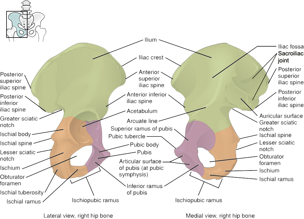
To get a comprehensive tour of these structures and their specific landmarks, we will use a detailed video.
Pelvis Hip Bones Anatomy (Os Coxae, Pelvic Girdle) - Ilium, Ischium, Pubis
The video "Pelvis Hip Bones Anatomy" from RegisteredNurseRN provides an excellent introduction to the os coxae. The presenter clearly explains the function of the pelvic girdle and introduces its three main regions.
Please watch the first part of the video from the beginning to the introduction. Pay attention to the overview of the three fused bones and the location of the acetabulum.
Now that you have a general understanding of the hip bone's composition, let's explore each of the three regions and their important bony landmarks in more detail.
The Ilium: The Flank Bone
The ilium is the largest and most superior of the three components, forming the "wings" of the pelvis that you can feel when you place your hands on your hips.
Pelvis Hip Bones Anatomy (Os Coxae, Pelvic Girdle) - Ilium, Ischium, Pubis
Let's continue with the same video to examine the ilium.
Watch the segment from the ilium. The video will walk you through its major landmarks. Focus on identifying the following structures: Iliac crest: The superior border you can palpate on your waist. Anterior Superior Iliac Spine (ASIS) and Anterior Inferior Iliac Spine (AIIS) on the front. Posterior Superior Iliac Spine (PSIS) and Posterior Inferior Iliac Spine (PIIS) on the back. Greater sciatic notch: The large notch on the posterior side. Auricular surface: The area that articulates with the sacrum.
To reinforce this information and connect it to a clinical context, please read the following text.
The Hip Bone - Ilium - Ischium - Pubis
This article from TeachMeAnatomy provides a concise summary of the ilium and highlights the clinical importance of the ASIS.
Read the section on the ilium, and pay close attention to the clinical relevance box about the Anterior Superior Iliac Spine. As a nursing student, you'll find that the ASIS is a critical landmark for many procedures and assessments, including measuring true leg length.
The Ischium: The "Sitting Bone"
The ischium forms the posteroinferior part of the hip bone. Its most prominent feature is the large, roughened area that supports your body weight when you sit down.
Pelvis Hip Bones Anatomy (Os Coxae, Pelvic Girdle) - Ilium, Ischium, Pubis
Now, let's move on to the ischium.
Watch the next segment of the video covering the ischium. As you watch, locate these key landmarks: Ischial tuberosity: The rough, thickened part you sit on. Ischial spine: A pointed projection that separates the greater and lesser sciatic notches. Lesser sciatic notch: A smaller notch inferior to the ischial spine. Ischial ramus: A branch of bone that projects anteriorly.
The ischial tuberosities are important clinical landmarks, especially in elderly or immobile patients, as they are common sites for pressure ulcers (bedsores).
The Pubis: The Anterior Bone
The pubis, or pubic bone, is the most anterior part of the hip bone. The two pubic bones meet in the midline at the pubic symphysis.
Pelvis Hip Bones Anatomy (Os Coxae, Pelvic Girdle) - Ilium, Ischium, Pubis
Finally, let's look at the pubis.
Watch the last major section of the video on the pubis. This part covers the following important features: Superior and Inferior pubic rami: The two branches that form part of a large opening. Pubic symphysis: The cartilaginous joint where the two pubic bones meet. Obturator foramen: The large hole formed by the ischium and pubis, which allows for the passage of nerves and blood vessels.
Now, let's read about the pubis and ischium for a quick review and to see another clinical connection.
The Hip Bone - Ilium - Ischium - Pubis
This reading will summarize the last two bones and provide a relevant clinical note on fractures.
First, read the section on The Pubis. Then, review the section on The Ischium. Note the clinical relevance box on Pubic Rami Fractures, which are common in elderly patients after low-impact falls.
Putting It All Together
You have now examined the three bones that form the os coxa and their key landmarks. To test your knowledge, take a look at this image of a real hip bone and try to identify the major features we've discussed without relying on the colors.

This practice is similar to what you might encounter in a lab setting with real bone models.
Conclusion
In this lesson, you've learned to identify the components and key landmarks of the pelvic girdle, or os coxae. We established that this structure is a fusion of three bones—the ilium, ischium, and pubis—and is built for stability and weight-bearing.
Key Takeaways:
- The os coxa (hip bone) is composed of the ilium, ischium, and pubis.
- These three bones fuse at the acetabulum, the socket for the hip joint.
- The ilium is the superior, fan-shaped bone, with key landmarks like the iliac crest and the anterior superior iliac spine (ASIS).
- The ischium is the posteroinferior bone, featuring the ischial tuberosity (the "sitting bone") and the ischial spine.
- The pubis is the anterior bone, which articulates with the opposing pubic bone at the pubic symphysis.
- The ischium and pubis together form the obturator foramen, the largest foramen in the skeleton.
You now have a solid foundation in the anatomy of the hip bone. In our next lesson, we will build on this knowledge to explore how the pelvic girdle differs structurally between males and females, an adaptation related to childbirth.