Welcome to the final lesson on the upper limb. Last time, we explored the forearm, identifying the radius and ulna and noting how their distal ends form the foundation for the wrist joint. We saw that the radius articulates directly with the wrist bones, playing a pivotal role in wrist movement.
Today, we will complete our journey down the appendicular skeleton's upper extremity by examining the intricate and fascinating structure of the hand and wrist. Our objective is to identify the bones of the wrist and hand: the carpals, metacarpals, and phalanges. The 27 bones in each hand allow for an incredible range of motion and dexterity, from powerful grips to delicate manipulations. For your future nursing practice, understanding this anatomy is crucial for assessing common injuries like fractures and conditions such as carpal tunnel syndrome, especially prevalent in the elderly patients you have experience with.
An Overview of the Hand's Skeleton
The bones of the hand are organized into three distinct groups. Let's start with a simple visual map to orient ourselves.
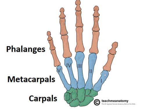
As you can see, the hand's structure progresses logically from the forearm:
- Carpals: A set of eight short bones that form the wrist.
- Metacarpals: Five long bones that form the palm of the hand.
- Phalanges: Fourteen long bones that constitute the fingers and thumb.
Now, let's dive into each of these groups in detail, starting with the most complex: the carpals.
The Carpal Bones: The Wrist
The wrist, or carpus, is a collection of eight irregularly shaped bones arranged in two rows: a proximal row that articulates with the radius and ulna, and a distal row that articulates with the metacarpals. Identifying these bones and their arrangement is a classic anatomy challenge.
The following video provides an excellent, detailed walkthrough of each carpal bone, its specific features, and its articulations.
Wrist & Hand Bones (Carpals, Metacarpals & Phalanges) | Anatomy
This video, "Wrist & Hand Bones" from Taim Talks Med, will guide you through the two rows of carpal bones, pointing out key landmarks on each one.
First, watch the detailed examination of the proximal row, starting from the lateral (thumb) side with the scaphoid and moving medially. Then, continue with the distal row, again moving from lateral to medial. The video provides a useful mnemonic to help you remember the order of the bones.
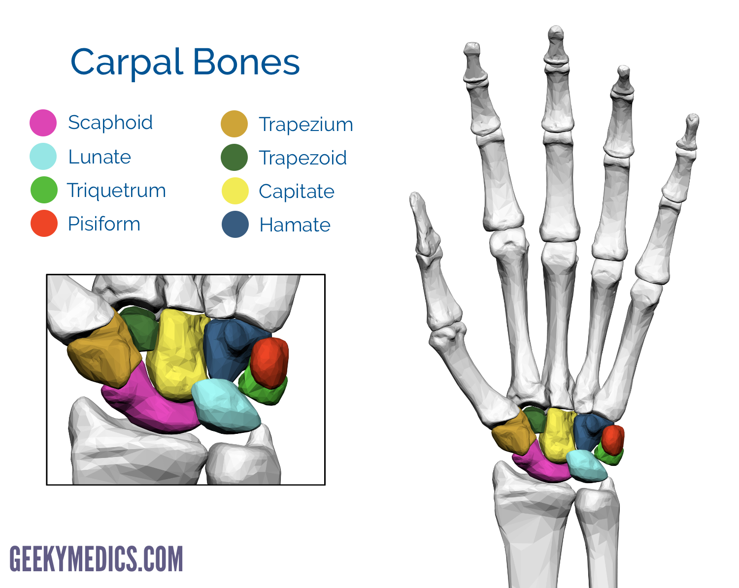
To summarize the arrangement from lateral (thumb side) to medial (pinky side):
- Proximal Row: Scaphoid, Lunate, Triquetrum, Pisiform
- Distal Row: Trapezium, Trapezoid, Capitate, Hamate
The mnemonic from the video is a great way to memorize this order: Scary Lions Try Pizza, That They Can't Handle.
Here is a color-coded diagram to help you visualize and memorize these eight bones.
To connect this anatomical knowledge to clinical practice, let's look at a resource that highlights a common injury.
The Bones of the Hand: Carpals, Metacarpals and Phalanges
This article from TeachMeAnatomy provides a concise written overview and includes important clinical context.
Please read the section on the carpal bones and pay special attention to the clinical relevance box discussing scaphoid fractures. This is the most commonly fractured carpal bone, often resulting from a fall on an outstretched hand (a "FOOSH" injury), something you might frequently encounter. Note the information about its "retrograde blood supply," which can lead to complications like avascular necrosis.
The TeachMeAnatomy article also mentions how the carpal bones form a U-shaped arch, which is enclosed by the flexor retinaculum to create the carpal tunnel. This anatomical space is a critical site for potential nerve compression, leading to carpal tunnel syndrome.
The Metacarpals and Phalanges: The Palm and Digits
Moving distally from the carpals, we find the bones that form the framework of the palm and fingers. These are more straightforward in their organization than the carpals.
Let's return to the "Taim Talks Med" video to walk through these bones.
Wrist & Hand Bones (Carpals, Metacarpals & Phalanges) | Anatomy
We will now complete our tour of the hand by looking at the metacarpals and phalanges.
Watch the segment on the metacarpals, noting their numbering and structure. Then, finish with the section on the phalanges, which explains the difference between the thumb and the other four fingers.
Metacarpals:
There are five metacarpals, numbered I to V, starting with the thumb (pollex). Each metacarpal consists of a proximal base (articulating with the distal carpal row), a shaft, and a distal head. The heads of the metacarpals form the knuckles of a clenched fist.
Phalanges:
These are the bones of the digits.
- Digits II through V (index, middle, ring, and little finger) each have three phalanges: a proximal, a middle (or intermediate), and a distal phalanx.
- The thumb (digit I) is an exception, having only two phalanges: a proximal and a distal phalanx.
This means there are 14 phalanges in total in each hand.
For a final review, let's quickly read through a textual summary of these two bone groups.
7.6C: Carpals, Metacarpals, and Phalanges (The Hand)
This article from LibreTexts provides a succinct description of the metacarpals and phalanges.
Read the short sections on the Metacarpals and the Phalanges to solidify your understanding of their numbering and structure.
Conclusion
In this lesson, we have successfully identified the 27 bones that comprise the wrist and hand. You are now able to distinguish between the three major groups and name the individual bones within each.
Key Takeaways:
- The hand contains carpals (wrist), metacarpals (palm), and phalanges (digits).
- The eight carpal bones are arranged in two rows:
- Proximal: Scaphoid, Lunate, Triquetrum, Pisiform
- Distal: Trapezium, Trapezoid, Capitate, Hamate
- The five metacarpals are numbered I-V, starting from the thumb.
- The phalanges are the finger bones. The thumb has two (proximal, distal), while the other four digits have three (proximal, middle, distal).
With this lesson, we conclude our study of the upper limb's appendicular skeleton. We have traced the path from the pectoral girdle all the way to the fingertips.
In our next session, we will shift our focus to the lower half of the body, beginning with the pelvic girdle. We will identify the bones that make up the pelvis and see how its structure is adapted for weight-bearing and locomotion.