Welcome to our next lesson on the biological and chemical foundations of neuroscience. In the previous lesson, we explored diffusion, the process by which ions and other particles move from an area of high concentration to one of low concentration. We established that this movement, driven by a concentration gradient, is a fundamental force shaping the cellular environment.
Today, we will introduce a second, equally important force: the electrical force. You'll learn how the interplay between concentration gradients and electrical forces gives rise to a crucial property of all neurons: the membrane potential. This is an electrical voltage across the cell membrane that acts like a charged battery, storing the energy the neuron will use to send signals. By the end of this lesson, you will be able to explain what electrical potential is in a biological context and how it is created and maintained.
From Batteries to Brains: What is Voltage?
Before we dive into the biology, let's establish a clear, intuitive understanding of what "voltage" or "electrical potential" really means. The term might bring to mind wall sockets or batteries, and that's actually a great place to start. In essence, voltage is a measure of potential energy that comes from a separation of electrical charges.
The video below from the "Be Smart" YouTube channel provides a fantastic historical and practical introduction to this idea. The host, Joe, explains how Alessandro Volta invented the first battery by separating charges using different metals. This is a perfect analogy for what happens in a neuron, just with different components.
How Your Brain Makes Its Own Electricity
This video, "How Your Brain Makes Its Own Electricity," offers a fun and intuitive look at bioelectricity. Please watch the segment where Joe builds a simple battery.
Focus on the explanation from Volta's experiments. He explains that voltage is the force driving charges (in his case, electrons) to move from one place to another. This separation of charges creates potential energy, just like in a stretched rubber band.
As the video explains, voltage is the potential for charges to move. In a battery, this potential is created by separating charges between two different metals. In a neuron, the same principle applies, but the components are different:
- The charge carriers are not electrons, but ions (like Na⁺ and K⁺).
- The barrier separating the charges is not air, but the cell's lipid membrane.
Let's formalize this connection.
Ion Gradients, Leak Channels, and the Na⁺/K⁺ Pump
The article "Ion Gradients, Leak Channels, and the Na⁺/K⁺ Pump" from Bio LibreTexts has a great section that directly translates the language of physics into the language of cell biology.
Please read the section titled Electricity and the Cell. Pay close attention to the table that maps physics terms like Voltage, Current, and Resistance to their biological meanings. The analogy of the dam is particularly helpful for visualizing this stored energy.
To summarize, the membrane potential is simply the voltage—the difference in electrical charge—between the inside and the outside of a neuron's membrane. This separation of charge turns the neuron into a tiny biological battery.
How a Neuron Builds its Charge
How does a neuron actually separate charges to create this voltage? The process is a beautiful interplay of the concepts we've learned so far: ions, a selectively permeable membrane, and concentration gradients.
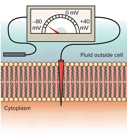
Scientists measure the membrane potential by placing a tiny electrode inside the cell and another one outside, as shown in the diagram below. By convention, the outside is considered the reference point (0 millivolts, or mV). In a typical resting neuron, the inside of the cell is about -70 mV relative to the outside. This is called the resting membrane potential.
Let's break down how this negative potential is generated step-by-step.
The membrane potential - Khan Academy
The Khan Academy article, "The membrane potential," provides a clear and detailed explanation of this process.
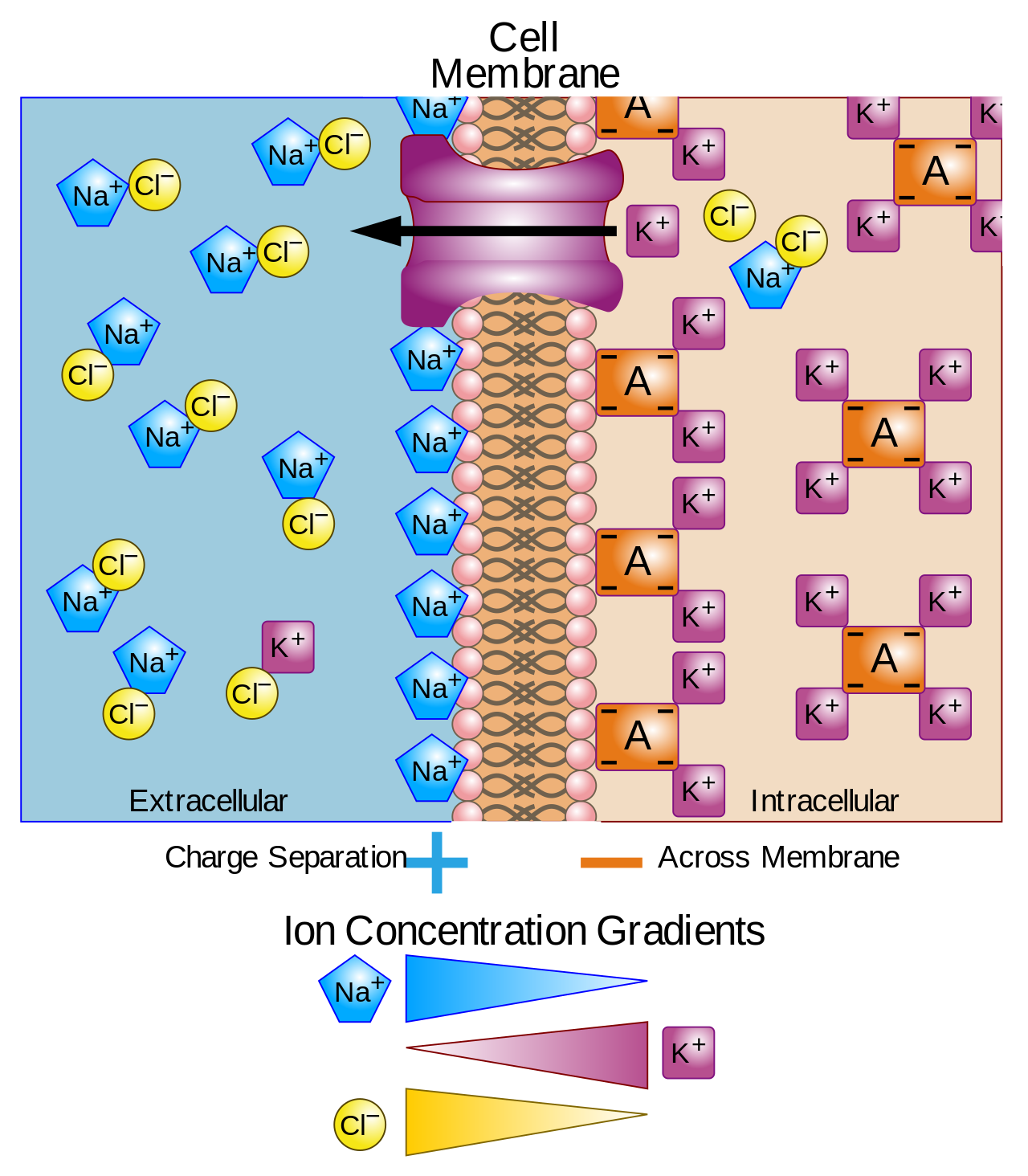
First, read the section defining the potential. This will formalize what we just discussed. Then, continue to the next section, "Where does the resting membrane potential come from?", which lists the key ions and their concentration differences across the membrane.
The Tug-of-War: Chemical vs. Electrical Forces
Now we get to the core of the concept. Imagine a simplified neuron where the membrane is only permeable to potassium (K⁺).
-
Chemical Force (Diffusion): As we learned in the last lesson, there is a high concentration of K⁺ inside the cell and a low concentration outside. This concentration gradient creates a chemical force that pushes K⁺ ions to diffuse out of the cell through their specific "leak" channels.
-
Creating an Electrical Force: Each K⁺ ion carries a single positive charge. As these positive charges leave the cell, they leave behind unbalanced negative charges inside the cell (mostly on large protein anions that are too big to leave). This creates a charge separation—and thus an electrical potential, or voltage—across themembrane. The inside becomes negative, and the outside becomes positive.
-
The Tug-of-War: This newly created electrical potential exerts its own force. Since opposite charges attract, the negative charge inside the cell now pulls the positive K⁺ ions back into the cell.
So, we have a "tug-of-war":
- The chemical force of the concentration gradient pushes K⁺ out.
- The electrical force of the membrane potential pulls K⁺ in.
The following animation masterfully illustrates this dynamic balance.
Membrane Potential, Equilibrium Potential and Resting Potential, Animation
The video "Membrane Potential, Equilibrium Potential and Resting Potential" by Alila Medical Media provides a fantastic animation of this process.
Watch the section from the beginning of the explanation. The video first introduces the two opposing forces and then animates how the movement of one ion type down its concentration gradient results in an electrical potential that opposes that very movement.
Finding the Balance: The Equilibrium Potential
What happens when this tug-of-war reaches a stalemate? The electrical force pulling K⁺ in eventually becomes strong enough to exactly balance the chemical force pushing K⁺ out. At this point, there is no net movement of K⁺. For every ion that leaves, another one enters. The system is in equilibrium.
The specific voltage at which this perfect balance is achieved is called the equilibrium potential for that ion.
For K⁺, the equilibrium potential is about -90 mV. This means that if the membrane potential were -90 mV, the electrical pull would be strong enough to perfectly counteract the powerful chemical push from its concentration gradient.
The Real World: From Equilibrium to Resting Potential
Of course, a real neuron membrane isn't just permeable to potassium. It is also slightly permeable to sodium (Na⁺) and chloride (Cl⁻). Each of these ions has its own concentration gradient and therefore its own equilibrium potential.
- Potassium (K⁺), as we've seen, has an equilibrium potential of about -90 mV. It "wants" to make the cell very negative.
- Sodium (Na⁺) is highly concentrated outside the cell. Its concentration gradient pushes it in. To balance this, the inside of the cell would need to be very positive to push it back out. Its equilibrium potential is about +60 mV. It "wants" to make the cell very positive.
The actual resting membrane potential of the neuron is the result of another tug-of-war, this time between the different ions. The final voltage will be a weighted average, settling closer to the equilibrium potential of the ion the membrane is most permeable to.
In a resting neuron, the membrane is about 50-100 times more permeable to K⁺ than to Na⁺. Therefore, K⁺ wins the tug-of-war by a large margin. The resting membrane potential of about -70 mV is much closer to K⁺'s equilibrium potential (-90 mV) than to Na⁺'s (+60 mV).
The membrane potential - Khan Academy
Let's return to the Khan Academy article to solidify these last two concepts. It walks through the single-ion and multi-ion scenarios in detail.
Read through the three sections starting with "What happens if only K⁺ can cross the membrane?" to see the step-by-step creation of the equilibrium potential. Then read the definition of equilibrium potential. Finally, read the section on the contribution of both ions, which explains the "tug-of-war" concept that determines the final resting potential.
Conclusion
In this lesson, we have combined our knowledge of ions, membranes, and gradients to understand one of the most fundamental properties of a neuron: its electrical potential.
Here are the key takeaways:
- Membrane potential (or voltage) is the separation of electrical charge across the cell membrane, which stores potential energy like a battery.
- It is created when ions move down their concentration gradient through a selectively permeable membrane.
- This ion movement separates charge, creating an opposing electrical gradient. The combined influence of these two forces is called the electrochemical gradient.
- The equilibrium potential is the voltage at which the electrical force exactly balances the chemical (concentration) force for a single ion type, resulting in no net movement.
- The resting membrane potential (~-70 mV) is the overall voltage in a resting neuron, determined by a "tug-of-war" between the equilibrium potentials of all permeable ions, weighted by their permeability. It is closest to the equilibrium potential for potassium (K⁺) because the membrane is most permeable to it.
We have established that the resting potential is a dynamic state, with K⁺ ions leaking out and Na⁺ ions leaking in. This raises a critical question: if ions are constantly leaking, wouldn't their concentration gradients eventually run down, causing the membrane "battery" to die? They would, if not for a special protein that works tirelessly to recharge it. In our next lesson, we will distinguish between the passive ion channels we've discussed so far and active ion pumps, focusing on the sodium-potassium pump, the machine that maintains the gradients essential for all neural activity.