Hello! Let's dive into the second lesson of our module on muscle physiology.
In our last lesson, we constructed the architectural blueprint of a skeletal muscle, journeying from the whole muscle down to the individual sarcomere. We identified the key protein filaments—actin and myosin—as the fundamental components of this microscopic engine.
Today, we transition from structure to function. If the sarcomere is the engine, the sliding filament theory is the operating manual that explains how it runs. We will unpack the precise, step-by-step process that allows these microscopic filaments to generate the force that moves your body.
Our learning outcome is to explain the sliding filament theory of muscle contraction in simple, functional terms. Understanding this process is not just an academic exercise; it's the foundation for comprehending why certain exercises feel harder at different points, how muscles generate force, and ultimately, how they are stimulated to grow.
1. From Intent to Action: Initiating a Contraction
Before the filaments can slide, they need a command. This command originates in your brain and travels as an electrical signal down a nerve to the muscle. The process of converting this electrical signal into a chemical "go" signal within the muscle cell is called excitation-contraction coupling.
Given your background in computer engineering, you can think of this as the initial API call that triggers a complex function. The brain sends the request, and a cascade of events unfolds to execute it at the hardware level.
This first video provides an excellent overview of this entire signaling chain, from the nerve impulse to the release of the key ingredient that starts the contraction: calcium.
Sliding Filament Model and Excitation Contraction Coupling
This video from Siebert Science, 'Sliding Filament Model and Excitation Contraction Coupling', will walk us through the 'big picture'—how a signal from the brain leads to the release of calcium within the muscle cell.
Watch the segment from 00:37 to 04:24. Focus on the sequence of events: Action Potential -> Neurotransmitter Release -> Signal travels down T-tubules -> Sarcoplasmic Reticulum (SR) releases calcium.
To summarize the key steps of this initiation phase:
- Signal Arrival: A nerve impulse (action potential) arrives at the neuromuscular junction (the synapse between nerve and muscle).
- Signal Propagation: The signal travels along the muscle fiber's membrane (sarcolemma) and down into the cell via the T-tubules.
- Calcium Release: The signal triggers the sarcoplasmic reticulum (SR)—the muscle cell's calcium reservoir—to release a flood of calcium ions (Ca²⁺) into the sarcoplasm, surrounding the myofilaments.
Calcium is the ultimate on-switch for muscle contraction. Without it, the machinery remains idle.
2. The Molecular Machinery: The Key Players
Now that calcium is present, the stage is set. Let's formally define the roles of the proteins involved in the contraction itself.
| Protein/Molecule | Role in Contraction | Analogy |
|---|---|---|
| Actin (Thin Filament) | The "track" along which myosin pulls. Contains binding sites for the myosin heads. | A rope or a ladder |
| Myosin (Thick Filament) | The "motor" protein. Its heads bind to actin and pull, generating force. | The hands pulling the rope |
| Tropomyosin | A regulatory protein that covers the myosin-binding sites on actin in a resting muscle. | A safety cover over the rungs of the ladder |
| Troponin | A regulatory protein attached to tropomyosin. It acts as the "lock" for the safety cover. | The lock on the safety cover |
| Calcium (Ca²⁺) | The "key" that binds to troponin, causing it to change shape and unlock tropomyosin. | The key that unlocks the lock |
| ATP | The "energy currency" that powers the myosin head for both pulling and detaching. | The energy supply for the person pulling the rope |
The interaction between these components is what constitutes the sliding filament theory.
3. The Cross-Bridge Cycle: The Algorithm of Contraction
With the players defined, let's examine the "algorithm" of muscle contraction. This process is a repeating sequence known as the cross-bridge cycle. Each cycle is a series of four main steps.
To visualize this process dynamically, let's watch a short, focused explanation.
How do Muscles Contract? Sliding Filament Theory | Corporis
The video 'How do Muscles Contract?' by Corporis provides a clear, animated breakdown of the cross-bridge cycle. It highlights the distinct roles of calcium and ATP in the process.
Watch the segment from 03:56 to 06:48. Pay close attention to how calcium exposes the binding sites and how ATP is used for both energy and detachment.
Let's break down that cycle step-by-step, using the excellent diagram below as a static reference.
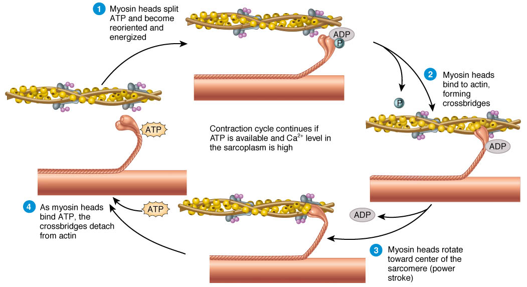
Here is the four-step cycle in detail:
-
Binding Site Exposure & Myosin Energizing:
- Calcium floods the cell and binds to troponin.
- This binding causes tropomyosin to shift, exposing the binding sites on the actin filament.
- Simultaneously, a myosin head hydrolyzes (breaks down) ATP into ADP and a phosphate group (Pi). This releases energy, which "cocks" the myosin head into a high-energy, ready position.
-
Cross-Bridge Formation (The "Grab"):
- The energized myosin head now attaches to the exposed binding site on the actin filament, forming what is called a cross-bridge.
-
The Power Stroke (The "Pull"):
- The myosin head releases the ADP and Pi. This release triggers the myosin head to pivot and bend forcefully.
- This pivot pulls the actin filament toward the M-line (the center of the sarcomere). This is the "power stroke"—the fundamental force-producing action of muscle contraction. The sarcomere shortens.
-
Detachment & Reset (The "Release"):
- A new ATP molecule binds to the myosin head.
- This binding causes the myosin head to detach from actin, breaking the cross-bridge.
- The new ATP is then hydrolyzed (back to step 1), re-energizing the myosin head to repeat the cycle.
This cycle of "grab, pull, release, reset" continues as long as calcium and ATP are present. Millions of these power strokes occurring in unison across thousands of sarcomeres result in the observable shortening of the entire muscle.
Muscle Relaxation occurs when the nerve signal ceases. Calcium is actively pumped back into the sarcoplasmic reticulum, troponin returns to its original shape, and tropomyosin slides back over the binding sites, preventing cross-bridges from forming.
4. From Theory to Training: The Length-Tension Relationship
This microscopic process has a direct and profound impact on your strength in the gym. The amount of force a muscle can produce is directly related to the number of cross-bridges it can form at any given moment. This relationship is determined by the overlap between actin and myosin filaments, which changes as the muscle lengthens and shortens. This is known as the length-tension relationship.
Sliding Filament Theory - Art of Anatomy
The article 'Sliding Filament Theory' from Art of Anatomy connects the microscopic theory to this crucial practical concept.
Read the final section titled 'Why does it matter?' and the subsection on the 'Length-tension relationship'.
Coach's Note: Optimal Overlap
As the reading explains, the sliding filament theory tells us:
- Mid-Range (Optimal Tension): When a muscle is in its mid-range of motion (neither fully stretched nor fully contracted), there is an optimal overlap of actin and myosin filaments. This allows for the maximum number of potential cross-bridges to be formed, resulting in the highest potential force output. This is why you are strongest in the middle of a bicep curl or bench press.
- Lengthened Position (Low Tension): When a muscle is fully stretched, the actin and myosin filaments are pulled far apart. There is minimal overlap, meaning fewer cross-bridges can form. This results in lower force-producing capacity.
- Shortened Position (Low Tension): When a muscle is fully contracted, the actin filaments from opposite ends of the sarcomere begin to overlap and interfere with each other, and the myosin filaments run up against the Z-discs. This "crowding" limits the number of effective cross-bridges that can form, again reducing force output.
Understanding this principle is fundamental to exercise selection and a topic we will revisit many times, particularly when discussing how to train muscles effectively through their entire contractile range.
Conclusion
Today we've demystified muscle contraction, translating it into a clear, logical sequence. You've learned that what appears as a smooth, simple movement is actually the sum of countless microscopic power strokes, all governed by a precise chemical and mechanical algorithm.
Key Takeaways:
- The sliding filament theory explains that muscle contraction occurs when myosin (thick) filaments pull actin (thin) filaments, shortening the sarcomere.
- The process is initiated by a nerve impulse that causes the release of calcium from the sarcoplasmic reticulum.
- Calcium acts as a key, binding to troponin to move tropomyosin and expose the binding sites on actin.
- The cross-bridge cycle is a four-step process: cross-bridge formation ("grab"), power stroke ("pull"), detachment ("release"), and reactivation ("reset").
- ATP is crucial for both energizing the power stroke and for detaching the myosin head from actin.
- The length-tension relationship, a direct result of filament overlap, explains why a muscle's strength varies throughout its range of motion.
In our next lesson, we will build upon this universal mechanism of contraction to explore the differences between muscle fiber types. We'll examine why some fibers are built for endurance (Type I) and others for explosive power (Type II), and what this means for our goal of maximizing hypertrophy.