Hello! Welcome to the second module of our course.
In the previous module, we established the foundational language of movement—anatomical terms, planes of motion, and joint actions. We finished by dissecting the dynamics of a single repetition, exploring how concentric, eccentric, and isometric contractions each play a unique and vital role in stimulating hypertrophy. We even got a sneak peek at the microscopic proteins involved, like actin, myosin, and the elastic protein titin.
Today, we transition from the dynamics of contraction to the structure of the contractor itself: the skeletal muscle. To truly understand how a muscle grows, we first need to understand what it's made of. This lesson zooms in, level by level, through the intricate architecture of a muscle.
Our learning outcome is to describe the hierarchical structure of a skeletal muscle, from the whole muscle down to the sarcomere.
Think of this like exploring the architecture of a complex computer system. You can't optimize the software without understanding the hardware it runs on. A muscle is a biological machine, and today, we're studying its schematic.
1. The Blueprint of a Muscle: Levels of Organization
A skeletal muscle isn't a single, uniform block of tissue. It's a highly organized, nested structure, where components are bundled within larger components. This hierarchical arrangement is key to its ability to generate and transmit immense force.
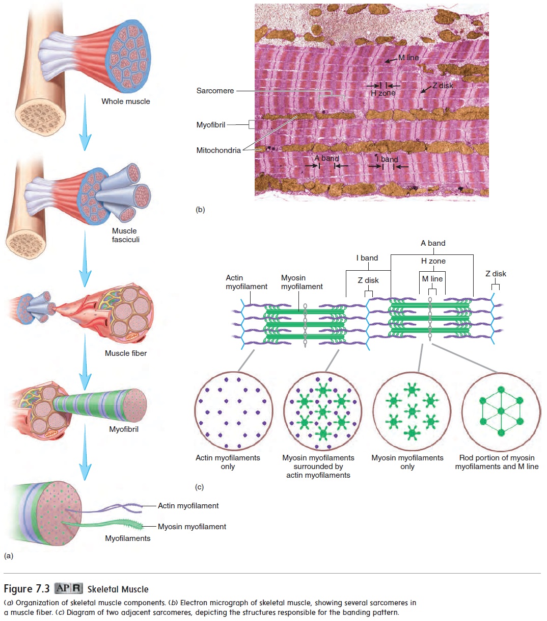
Let's start with a visual overview. The image below illustrates this "nested doll" structure, which we will be breaking down piece by piece.
To walk through these levels, we'll watch a clear, concise video that explains each layer of the hierarchy.
Skeletal Muscle Levels of Organization + Filament Basics
This video from Siebert Science, 'Skeletal Muscle Levels of Organization', provides an excellent step-by-step breakdown from the whole muscle down to the molecular level. It introduces the core terminology we'll use throughout the course.
Watch the video from the beginning to 06:18. Focus on memorizing the name and order of the five levels of organization presented. The video repeats them multiple times, which is helpful for retention.
As the video detailed, the hierarchy is as follows:
| Level of Organization | Structure | Description |
|---|---|---|
| Organ | Muscle | The complete muscle (e.g., biceps brachii). |
| Tissue | Fascicle | A bundle of muscle fibers. |
| Cellular | Muscle Fiber | A single, elongated, multinucleated muscle cell. |
| Organelle | Myofibril | A rod-like contractile organelle within a muscle fiber. |
| Molecular | Myofilaments | The protein filaments (actin and myosin) that make up the myofibril. |
2. The Connective Tissue Harness: Transmitting Force
The video gave us the "what," but it's also crucial to understand the "how." How do these nested bundles work together? The answer lies in the connective tissue that encases each level. This isn't just wrapping; it's a structural harness that funnels the force generated by microscopic proteins into meaningful movement of your skeleton.
To understand this, please read the first part of the following resource.
8.2 Skeletal Muscle – Human Physiology
This text from the 'Human Physiology' textbook by LMU Pressbooks clearly describes the three layers of connective tissue that organize and transmit force through the muscle.
Read the first section, which ends just before the 'Skeletal Muscle Fibers' heading. Pay close attention to the names and locations of the three 'mysia' layers: epimysium, perimysium, and endomysium.
Let's summarize these crucial layers:
- Epimysium ("upon the muscle"): A dense sheath of connective tissue that covers the entire muscle.
- Perimysium ("around the muscle"): Connective tissue that bundles muscle fibers into fascicles.
- Endomysium ("within the muscle"): A delicate layer of connective tissue that surrounds each individual muscle fiber.
Think of this system like a rope. Thousands of tiny fibers (muscle fibers) are bundled into yarns (fascicles), which are then twisted together to form the final, strong rope (the whole muscle). The force from each tiny fiber is transmitted through the endomysium, to the perimysium, to the epimysium, and finally into the tendon, which pulls on the bone. An injury to this connective tissue can compromise the muscle's ability to transmit force, even if the muscle fibers themselves are healthy.
3. The Muscle Fiber: The Site of Growth
Now we zoom into the cellular level—the muscle fiber (or myofiber). This is the actual cell that undergoes hypertrophy. It's a unique and highly specialized cell.
As you learned in the video, a muscle fiber contains specialized organelles. Let's revisit the key ones:
- Sarcolemma: The cell membrane of the muscle fiber.
- Sarcoplasm: The cytoplasm of the muscle fiber.
- Sarcoplasmic Reticulum (SR): A network that stores, releases, and retrieves calcium ions—the "on switch" for muscle contraction.
- Transverse Tubules (T-tubules): Inward folds of the sarcolemma that allow electrical signals to travel deep into the cell.
Coach's Note: Satellite Cells
For our purposes, there's another critical component associated with the muscle fiber that is the key to long-term growth: the satellite cell. This is one of the most important concepts for understanding how muscles adapt and grow.
8. Structure and Function of Exercising Muscle
This reading from 'Structure and Function of Exercising Muscle' introduces the concept of satellite cells, which are fundamental to muscle repair and hypertrophy.
Please read the section titled 'Muscle Fibers'. Focus on the description and function of satellite cells.
As the text explains, satellite cells are myogenic (muscle-generating) stem cells that lie dormant on the surface of the muscle fiber. When you train and cause micro-damage to the muscle, these cells are activated. They multiply and can donate their nuclei to the existing muscle fiber.
Why is this important? The nucleus is the "command center" of the cell, directing protein synthesis. A single nucleus can only manage a certain volume of cytoplasm (this is called its myonuclear domain). For a muscle fiber to grow larger (hypertrophy), it needs more nuclei to manage the increased cellular machinery. Satellite cells provide these new nuclei, enabling the muscle fiber to synthesize more contractile proteins and expand its size. This is the biological engine of muscle growth.
4. The Sarcomere: The Engine of Contraction
Inside each muscle fiber are thousands of myofibrils. And each myofibril is a long chain of repeating functional units called sarcomeres. The sarcomere is the smallest and most fundamental contractile unit of a muscle. It's where the magic of contraction happens.
The distinct, striped (or "striated") appearance of skeletal muscle comes from the highly organized arrangement of proteins within the sarcomere. Let's define these components.
8.2 Skeletal Muscle – Human Physiology
We'll return to the 'Human Physiology' text to get a clear definition of the sarcomere and its constituent parts.
Read the section titled 'The Sarcomere'. Focus on understanding the definitions of the Z-disc, A-band, I-band, and H-zone, and which myofilaments are present in each.
Referring back to our main diagram, let's crystallize these key parts of the sarcomere:
- Myofilaments: The actual contractile proteins.
- Thick Filaments: Made of the protein myosin.
- Thin Filaments: Made primarily of the protein actin.
- Z-Discs: The boundaries at each end of a sarcomere, where the thin filaments are anchored. A sarcomere is defined as the area from one Z-disc to the next.
- Bands & Zones: These describe the appearance of the sarcomere based on filament overlap.
- A-Band: The dark region containing the entire length of the thick (myosin) filaments.
- I-Band: The light region containing only thin (actin) filaments.
- H-Zone: The central, slightly lighter area within the A-band, containing only thick (myosin) filaments.
When a muscle contracts, these myofilaments slide past each other, causing the Z-discs to move closer together and shortening the entire sarcomere. When millions of sarcomeres shorten in unison, the entire muscle contracts.
Conclusion
Today, we've journeyed from the macroscopic muscle you see in the mirror down to the microscopic engine that powers it. This hierarchical structure is a marvel of biological engineering, designed for efficient force production and transmission.
Key Takeaways:
- Skeletal muscle has a nested hierarchy: Muscle → Fascicles → Muscle Fibers → Myofibrils → Sarcomeres.
- Connective tissue layers (epimysium, perimysium, endomysium) provide structural integrity and transmit force from the fibers to the tendons.
- The muscle fiber is the individual muscle cell that grows, a process aided by the donation of new nuclei from satellite cells.
- The sarcomere is the basic contractile unit, composed of interdigitating actin (thin) and myosin (thick) filaments. Its structure gives muscle its striated appearance.
You now have the anatomical blueprint. You know the names of the parts and their arrangement. In our next lesson, we will bring this blueprint to life by exploring the sliding filament theory, which describes exactly how actin and myosin interact to generate force and cause muscle contraction.