Introduction
Welcome back. In our previous lesson, we established the "why" and "how" of movement screening, focusing on using tools like the Overhead Squat Assessment (OHSA) to identify compensations. You learned to spot deviations from ideal movement, such as knee valgus or an excessive forward lean, and link them to a general list of overactive and underactive muscles.
Today, we transition from observation to diagnosis. This lesson is designed to help you master the learning outcome: Diagnose common movement faults (e.g., knee valgus in a squat) by linking them to specific muscular imbalances or mobility restrictions.
Think of this as building a diagnostic algorithm. When a client performs a movement, they provide you with data. A fault, like their knee caving in, is an error message. Your job is to trace that error back to its source code—the specific muscular or mobility issue causing the problem. This process of observing, hypothesizing, and testing is where your anatomical knowledge transforms into a powerful coaching tool that delivers superior results and builds profound client trust.
1. Diagnosing Lower Body Movement Faults: The Squat
The squat is a foundational pattern that reveals a great deal about a client's movement capabilities. We'll use it as our primary model for diagnosis. A fault is rarely caused by a single issue; it's often a combination of factors. Your task is to determine the primary driver.
Fault 1: Knee Valgus ("Knee Cave")
Knee valgus is the inward collapse of the knees, especially during the ascent of a squat. While it can occur on the way down, it's most often a sign that the body is struggling to produce force on the way up.
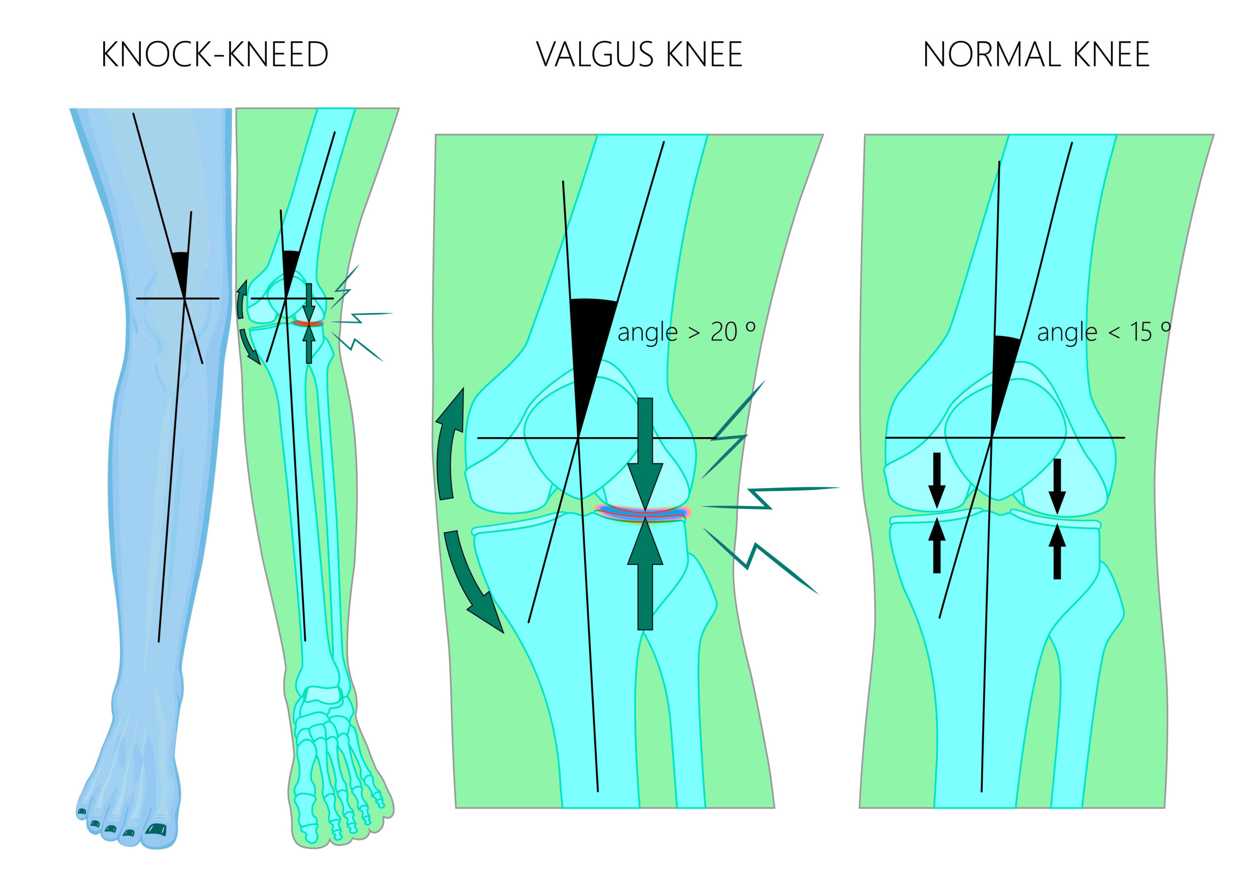
To diagnose the cause, we follow a logical sequence, starting from the ground up.
Knee Valgus Explained: Fix Knee Cave and Boost Your Strength
Dr. Aaron Horschig from Squat University provides an excellent framework for understanding knee valgus. He emphasizes that it's primarily a stability issue. Let's watch how he breaks down the diagnostic process.
Watch the following segments: 00:00 - 01:45: Pay close attention to the concept of the 'tripod foot.' A collapse of the foot's arch is often the first domino to fall, leading to knee valgus. 03:24 - 04:24: Observe how the single-leg squat is used as a specific test for dynamic knee control. Wobbling or an immediate knee cave during this test points directly to a lack of stability.
Diagnostic Algorithm for Knee Valgus:
-
Check the Foot (The Foundation):
- Observation: Watch the client's feet during a bodyweight squat. Does the arch of the foot collapse inward (over-pronation)? Does the weight shift away from the outside of the foot?
- Hypothesis: If the foot collapses, the issue could be a lack of motor control to maintain the "tripod foot" or a mobility restriction elsewhere (like the ankle) that forces the foot to compensate.
- Underlying Cause: This points to underactive intrinsic foot muscles and potentially overactive peroneal muscles on the outside of the lower leg.
-
Check the Ankle (Mobility):
- Observation: Does the client have difficulty squatting deep without their heels lifting or their torso leaning excessively forward?
- Hypothesis: As detailed in the resource "5 Common Squat Compensations," impaired ankle dorsiflexion can cause the foot to pronate and the knee to subsequently cave in as the body searches for range of motion.
- Test: Use the Knee-to-Wall Test from our last lesson. A score of less than 10-12 cm is a strong indicator of an ankle mobility deficit.
-
Check the Hips (Strength & Control):
- Observation: The knee caves in, but the foot and ankle appear stable.
- Hypothesis: This is the classic presentation of weak hip abductors and external rotators, namely the gluteus medius and gluteus maximus. These muscles are responsible for pulling the femur outward and preventing it from collapsing inward. Overactive adductors (inner thigh muscles) can also contribute by actively pulling the knee in.
- Test: The single-leg squat is your go-to diagnostic tool here. If the client cannot maintain knee alignment on one leg, it confirms a deficit in dynamic hip stability.
| Observation | Primary Hypothesis | Likely Imbalance |
|---|---|---|
| Knee caves inward + foot arch collapses. | Poor foot motor control or ankle mobility restriction. | Underactive intrinsic foot muscles; potential ankle dorsiflexion limitation. |
| Knee caves inward but foot remains stable. | Weak hip stabilizers. | Underactive gluteus medius/maximus; potentially overactive adductor complex. |
| Knee valgus appears only under heavy load/fatigue. | Strength deficit in hip stabilizers. | The gluteus medius/maximus are strong enough for light loads but fail under high demand. |
Fault 2: "Butt Wink" (Posterior Pelvic Tilt)
The "butt wink" is when the pelvis tucks under at the bottom of the squat, causing the lumbar spine to flex or round. A small amount of tuck at the absolute end range is normal, but a significant wink occurring before reaching full depth is a compensation.
5 Common Squat Compensations - Bull City Physical Therapy
The article '5 Common Squat Compensations' clearly outlines the primary causes of the butt wink. Let's review its diagnostic criteria.
Read the section 'Compensation #1: The “Butt Wink”'. Focus on the three potential causes it lists: ankle mobility deficits, hip mobility deficits, and decreased lumbopelvic stability.
Diagnostic Algorithm for Butt Wink:
- Test Ankle Mobility: As with knee valgus, limited ankle dorsiflexion is a primary culprit. If the knees can't travel forward enough, the pelvis must tuck under to allow the torso to descend further.
- Test: Use the Knee-to-Wall test. Also, try having the client squat with their heels on small plates. If the butt wink disappears, you have very likely found your primary cause.
- Test Hip Mobility: The hip socket has a finite amount of flexion. Once you reach that limit, the pelvis will rotate backward to continue the movement.
- Test: Have the client lie on their back and pull one knee to their chest (passive hip flexion). Observe the point at which their low back starts to press firmly into the floor or their tailbone begins to lift. If this happens well before the thigh contacts the chest, a structural or soft-tissue limitation in hip flexion may be the cause.
- Test Core Stability (Motor Control): The butt wink can also be a stability problem, where the client lacks the motor control to keep their pelvis neutral as they move into deep hip flexion.
- Test: Have the client perform a goblet squat. The weight held in front acts as a counterbalance and often cleans up the pattern by forcing more core engagement. If the butt wink improves significantly, it points to a motor control issue.
Fault 3: "Good Morning" Squat (Hips Rise First)
This fault occurs during the ascent from the bottom of the squat, where the hips shoot up much faster than the chest, causing the torso to pitch forward.
Diagnostic Algorithm for "Good Morning" Squat:
As the "5 Common Squat Compensations" article notes, this is almost always due to one of two things:
- Quadriceps Strength Deficit: The client's posterior chain (glutes, hamstrings) is significantly stronger than their quadriceps. To lift the weight, their body shifts the movement into a hip hinge pattern to leverage the stronger muscles, effectively turning the squat into a good morning. This is especially common under heavy loads.
- Motor Control Issue: The client simply has a flawed movement pattern and doesn't know how to properly coordinate knee and hip extension simultaneously.
- Test: Observe the squat at different loads. If the fault only appears at near-maximal weights, it strongly indicates a quad strength deficit. If it happens even with an empty bar, it may start as a motor control issue that can be corrected with cueing and lighter, deliberate practice.
2. Diagnosing Upper Body Movement Faults
Faults in pressing and pulling often stem from poor scapular control and thoracic spine mobility.
Fault 1: Winged Scapula
Scapular winging is when the medial (inner) border of the shoulder blade lifts away from the ribcage. It is most apparent during pushing movements like push-ups or an overhead press.

Diagnostic Algorithm for Winged Scapula:
- Observation: Have the client perform a wall push-up or hold a plank. Watch their shoulder blades as they press away from the wall or floor. If the inner edge of one or both scapulae becomes prominent, you have identified winging.
- Primary Cause: This is a classic sign of weakness or poor activation of the Serratus Anterior. This muscle's job is to protract the scapula (wrap it around the ribcage). When it's not doing its job, the scapula becomes unstable.
Fault 2: Shoulder Elevation/Rounding in Rows & Pull-ups
This fault occurs when a client initiates a pull by shrugging their shoulders up towards their ears (elevation) or allows their shoulders to roll forward (protraction) at the peak contraction of a row.
5 Upper-Body Training Mistakes - Advanced Human Performance
The article '5 Upper-Body Training Mistakes' provides an excellent analysis of this exact problem. It explains why simply 'pulling as far as possible' is often incorrect and leads to dysfunction.
Read sections '1. Over-Pulling On Pull-ups and Pulldowns' and '3. Rowing With Excessive ROM'. Pay attention to how excessive range of motion disengages the target muscles (lats, rhomboids) and shifts the work to the upper traps and shoulders, causing faulty mechanics.
Diagnostic Algorithm for Faulty Pulling Mechanics:
- Observation: During a lat pulldown or row, watch the client's shoulders. Do they shrug up at the beginning of the pull? Do they round forward at the end to try and get more range of motion?
- Primary Cause: This is a motor control problem. The client is dominant in their upper trapezius and levator scapulae, and they are under-utilizing the muscles that should be driving the movement: the latissimus dorsi (for shoulder depression and extension) and the mid/lower trapezius and rhomboids (for scapular retraction and depression).
- Test: Use a simple coaching cue. Ask the client to think about "putting their shoulder blades in their back pockets" or "driving their elbows down and back." If they can correct the pattern with this cue, it confirms a motor control deficit, not a structural limitation.
Conclusion
You have now moved beyond simple observation into the realm of diagnostics. By systematically assessing movement, you can build a strong hypothesis about the root cause of a fault—whether it's poor mobility, a lack of stability, a specific strength deficit, or a flawed motor pattern. This analytical approach, much like debugging code, allows you to be precise and effective.
Key Takeaways:
- Think Like a Detective: Start with the observed fault (e.g., knee valgus) and work backward using a logical sequence of tests to isolate the cause.
- Lower Body Diagnosis: For squats, always check the feet and ankles first, then move up to the hips and core. Common faults like knee valgus, butt wink, and the "good morning" squat can often be traced to predictable mobility or stability issues.
- Upper Body Diagnosis: For pushing and pulling, focus on scapular control. Faults like scapular winging and shoulder elevation are almost always signs of poor motor control and muscular imbalance (e.g., weak serratus anterior, overactive upper traps).
- Test Your Hypothesis: Use simple tests (e.g., single-leg squat, knee-to-wall test, goblet squat, corrective cueing) to confirm or deny your initial hypothesis about a fault's cause.
Preview of the Next Lesson
Diagnosing a problem is step one. Step two is solving it. What if a client's mobility or limb length makes a standard exercise unsafe or ineffective for them? In our next lesson, we will focus on application: you will learn to modify exercises for clients with different limb lengths and mobility restrictions using anatomical and biomechanical reasoning. This will equip you to truly individualize training for anyone who walks into the gym.