Hello! Welcome to the fourth lesson in our exploration of the foundations of neural communication.
In our last lesson, we saw how a neuron sends a signal across a synapse by releasing chemical messengers called neurotransmitters. We ended with the neurotransmitter binding to a receptor on the postsynaptic neuron. But what happens next? Does this always cause the next neuron to fire?
The answer is no. A signal can be a "Go!" or a "Stop!" message. Today, we'll explore this critical distinction. We will learn how neurotransmission can be either excitatory, making a neuron more likely to fire, or inhibitory, making it less likely to fire.
Understanding this balance is fundamental to neuroscience. The brain isn't just a chain of dominoes falling one after another; it's a sophisticated computational device that constantly weighs excitatory and inhibitory inputs to process information and make decisions. This balance is essential for everything from generating a thought to executing a smooth, controlled movement—a process that, as we'll see, goes awry in Parkinson's disease.
Excitatory vs. Inhibitory: "Go!" or "Stop!"
When a neurotransmitter binds to a postsynaptic receptor, it causes a small, temporary change in the membrane potential of the receiving neuron. This is called a postsynaptic potential (PSP). These potentials come in two flavors.
Excitatory and Inhibitory Postsynaptic Potentials
To start, let's get a formal definition of these two types of signals. This short text from the online textbook Neuroscience (2nd ed.) provides a clear and concise introduction.
Please read the first two paragraphs of this page. Focus on how Excitatory Postsynaptic Potentials (EPSPs) and Inhibitory Postsynaptic Potentials (IPSPs) are defined based on their effect on the neuron's likelihood to fire an action potential.
As the text explained, the distinction is simple and functional:
- An Excitatory Postsynaptic Potential (EPSP) increases the probability that the postsynaptic neuron will fire an action potential. It's a "Go!" signal.
- An Inhibitory Postsynaptic Potential (IPSP) decreases the probability that the postsynaptic neuron will fire an action potential. It's a "Stop!" signal.
The "decision" to fire an action potential is made at the axon hillock, where the neuron sums up all the incoming signals. An action potential is triggered only if the membrane potential at the axon hillock reaches the threshold of excitation (typically around -55 mV).
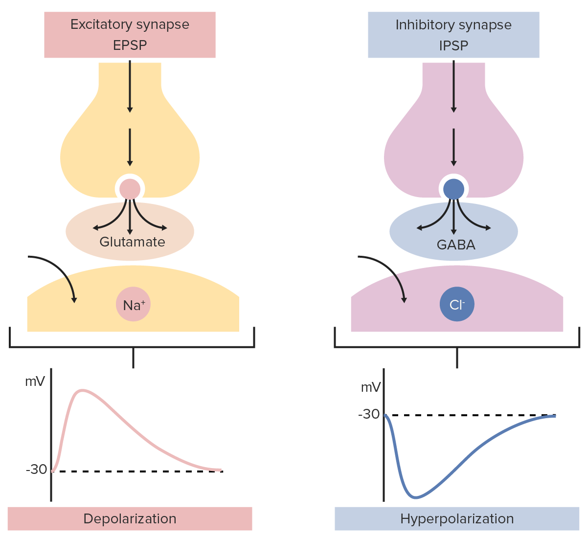
- EPSPs cause the membrane to depolarize (become less negative), moving the potential closer to the threshold.
- IPSPs typically cause the membrane to hyperpolarize (become more negative), moving the potential further away from the threshold.
The image below provides a great visual summary of the two processes.
Now, let's look at the specific ions and neurotransmitters that create these opposing effects.
The Mechanism of Excitation
The primary excitatory neurotransmitter in the brain is glutamate. When glutamate is released into a synapse, it binds to receptors on the postsynaptic neuron that are typically linked to ion channels permeable to positive ions.
To see how this works, let's watch a video that breaks it down.
Synaptic Transmission | How Neurons Communicate
The following video from 'Dr Matt & Dr Mike' clearly explains the ionic basis of both excitatory and inhibitory signals.
Please watch from 05:07 to 06:46. This segment focuses on how excitatory neurotransmitters work by opening specific ion channels. Then, watch the summary from 07:20 to 09:03, which recaps both types of transmission.
As the video explained, the process for an EPSP is straightforward:
- An excitatory neurotransmitter like glutamate binds to its ionotropic receptor.
- The receptor, which is a channel, opens.
- This channel is primarily permeable to sodium ions ().
- Since there's a high concentration of outside the cell, these positively charged ions rush into the neuron.
- This influx of positive charge makes the inside of the cell less negative, causing a small depolarization—an EPSP.
If enough of these EPSPs occur close together in time and space, they can sum up to push the neuron's membrane potential to the threshold, triggering an action potential.
The Mechanism of Inhibition
Just as crucial as the "Go!" signals are the "Stop!" signals that prevent the brain from becoming over-excited. Inhibition provides the control and sculpting necessary for precise neural processing. The main inhibitory neurotransmitter in the brain is GABA (gamma-aminobutyric acid). In the spinal cord, glycine also plays a major inhibitory role.
Let's return to the same video to see how these inhibitory signals work.
Synaptic Transmission | How Neurons Communicate
Now let's examine the other side of the coin: inhibition. This segment from the same 'Dr Matt & Dr Mike' video explains the ionic movements that make a neuron less likely to fire.
Please watch from 06:28 to 07:20. Pay attention to the two different ways that inhibition can be achieved through the movement of chloride and potassium ions.
Inhibition works by making the inside of the neuron more negative, or clamping it at its negative resting state, thus moving it further from the action potential threshold. This is achieved in one of two primary ways:
-
Chloride Ion () Influx: The inhibitory neurotransmitter (e.g., GABA) binds to its ionotropic receptor, opening a channel permeable to negatively charged chloride ions (). The influx of these negative ions makes the cell's interior more negative, causing hyperpolarization—an IPSP.
-
Potassium Ion () Efflux: Some inhibitory receptors, when activated, open channels permeable to potassium ions (). Since is more concentrated inside the neuron, these positive ions flow out of the cell. The loss of positive charge also makes the cell's interior more negative, resulting in hyperpolarization and an IPSP.
Both mechanisms make it harder for the neuron to reach the threshold for firing an action potential.
Test your understanding!
A certain drug is known to block GABA receptors. If this drug is administered, what would be the likely effect on the overall electrical activity in the brain? Would neurons become more or less likely to fire action potentials? Why?
Show answer
By blocking GABA receptors, the drug prevents the brain's main inhibitory neurotransmitter from working. This means that the "Stop!" signals are effectively silenced. As a result, the balance shifts heavily towards excitation. Neurons would become much more likely to fire action potentials, leading to uncontrolled, widespread electrical activity. This is the mechanism behind some types of seizures.
Neural Integration: The Neuron as a Calculator
A typical neuron in your brain is not just listening to one other neuron. It's receiving thousands of inputs at once—some excitatory, some inhibitory—at synapses all over its dendrites and cell body.
The neuron must somehow integrate this barrage of information to make a single, binary decision: fire an action potential, or stay quiet. This process is called summation.

The neuron continuously adds up all the EPSPs (depolarizations) and subtracts all the IPSPs (hyperpolarizations). If the resulting net change in voltage at the axon hillock reaches the threshold, an action potential is generated. If not, the neuron remains silent.
Given your interest in computer science, you can think of the neuron as a tiny biological processor. It takes multiple analog inputs (the varying strengths of EPSPs and IPSPs) and performs a computation (summation) to produce a single digital output (the all-or-none action potential). This constant, dynamic balance between excitation and inhibition is the fundamental basis of computation in the nervous system.
For more detail on the specific receptors involved and where these different synapses tend to occur on the neuron, this chapter from Neuroscience Online is an excellent resource.
Please read section '13.2 Spatial and Morphological Distinctions' to see how excitatory and inhibitory synapses are often located in different places on the neuron. Then, skim sections '13.10 Glutamate Receptors' and '13.11 Receptors-GABAA and Glycine' to reinforce which ions are associated with each type of neurotransmission.
Conclusion
Today, we've explored the crucial difference between the brain's "Go!" and "Stop!" signals. This balance is not just an abstract concept; it's the very foundation of how our brains function, moment to moment.
Key takeaways from this lesson:
- Synaptic transmission can be excitatory (EPSP), making a neuron more likely to fire, or inhibitory (IPSP), making it less likely to fire.
- Excitation is primarily mediated by the neurotransmitter glutamate, which opens channels for positive ions (like ) to enter the cell, causing depolarization.
- Inhibition is primarily mediated by GABA (and glycine), which opens channels for negative ions () to enter or positive ions () to exit, causing hyperpolarization.
- Neurons perform summation, integrating thousands of EPSPs and IPSPs to "decide" whether to fire an action potential.
We've now seen how a signal is initiated, transmitted across the synapse, and interpreted as either excitatory or inhibitory. But for the system to work effectively, the signal can't linger forever. The neurotransmitter must be cleared from the synapse to prepare for the next signal.
In our next lesson, we will examine the "clean-up" mechanisms: neurotransmitter reuptake and degradation. These processes are not only vital for normal brain function but are also the targets of many medications, including some used in the context of Parkinson's disease.