Hello! Welcome to your third lesson in our journey through the neuroscience of Parkinson's disease.
In our last lesson, we traced the journey of the action potential—the neuron's electrical signal—as it travels down the axon. We ended with a crucial question: What happens when this signal reaches the end of the line? How does one neuron "talk" to the next?
Today, we will answer that question by exploring the synapse, the remarkable junction where neural communication occurs. You will learn how the electrical signal of an action potential is converted into a chemical signal that bridges the gap between neurons. This process, known as synaptic transmission, is fundamental to all brain functions, from thinking and feeling to moving.
Specifically, we'll focus on how chemical messengers called neurotransmitters are released from one neuron and bind to specialized receptors on another. Understanding this process is essential, as the entire story of Parkinson's disease revolves around a disruption in this system, particularly involving the neurotransmitter dopamine.
The Synapse: The Brain's Communication Hub
A synapse is a highly specialized structure that allows a neuron (the presynaptic neuron) to pass a signal to another cell (the postsynaptic neuron). In the vast majority of cases in the central nervous system, this is not a direct electrical connection. Instead, there's a tiny gap called the synaptic cleft, which the signal must cross using a chemical messenger.
This entire process can be broken down into a few key steps, as illustrated in the diagram below.
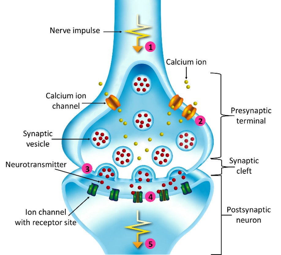
The core idea is a conversion of energy:
Electrical Signal (Action Potential) → Chemical Signal (Neurotransmitter) → Electrical Signal (Postsynaptic Potential)
To make this process intuitive, you can think of it using a simple analogy:
- The presynaptic terminal is like a mailroom with prepared packages.
- The neurotransmitters are the letters inside those packages.
- The synaptic vesicles are the packages themselves.
- The synaptic cleft is the space between the mailroom and the recipient's mailbox.
- The postsynaptic receptors are like the recipient's mailbox, shaped to accept only specific letters.
Let's now dive into the detailed biological mechanisms that make this elegant communication possible.
Step 1: From Electrical Impulse to Chemical Release
The process begins when the action potential we studied in the last lesson completes its journey along the axon and arrives at the axon terminal. This arrival is the trigger that initiates the entire sequence of chemical release.
To see exactly how this happens, please watch the following segment from a video by EKG Science. It clearly explains the critical role of calcium ions and the machinery involved in releasing neurotransmitters.
Synapses (Transmission): Electrical & Chemical | Neurology
This video, 'Synapses (Transmission): Electrical & Chemical', will walk us through the molecular steps of neurotransmitter release.
Please watch from 05:07 to 08:35. Pay close attention to the sequence of events starting with the action potential's arrival and ending with the release of neurotransmitters.
As the video detailed, the release of neurotransmitters is a precisely orchestrated event:
- Arrival of Action Potential: The wave of depolarization from the action potential reaches the presynaptic terminal.
- Opening of Calcium Channels: This depolarization opens special voltage-gated calcium () channels.
- Calcium Influx: Because there is a much higher concentration of outside the neuron than inside, these ions flood into the axon terminal.
- Vesicle Fusion: The influx of acts as a crucial signal. It binds to proteins (like synaptotagmin) associated with synaptic vesicles. This triggers a conformational change in a complex of proteins called SNAREs, which act like molecular ropes, pulling the vesicle to the presynaptic membrane and forcing it to fuse.
- Exocytosis: The fusion of the vesicle with the membrane causes it to spill its contents—the neurotransmitter molecules—into the synaptic cleft. This process is called exocytosis.
This entire sequence, from the action potential's arrival to neurotransmitter release, is incredibly fast, taking less than a millisecond.
Test your understanding!
Imagine a hypothetical toxin that prevents voltage-gated channels at the axon terminal from opening. A neuron affected by this toxin is still able to generate and propagate an action potential perfectly down its axon. What would happen when that action potential reaches the terminal? Would the neuron be able to communicate with the next one?
Show answer
Even though the action potential arrives successfully, the toxin-blocked channels would not open. Without the influx of calcium, the synaptic vesicles would not be triggered to fuse with the presynaptic membrane. Therefore, no neurotransmitter would be released into the synaptic cleft, and the signal would not be passed to the postsynaptic neuron. Communication would fail at the synapse.
Step 2: Crossing the Cleft and Binding to Receptors
Once released, the neurotransmitter molecules diffuse across the tiny synaptic cleft and bind to receptor proteins embedded in the membrane of the postsynaptic neuron. This binding is highly specific, often described using a "lock and key" model: a specific neurotransmitter (the key) will only fit into a specific type of receptor (the lock).
The effect of the neurotransmitter depends entirely on the type of receptor it binds to. There are two main families of receptors.
Synapses (Transmission): Electrical & Chemical | Neurology
Let's return to the 'Synapses (Transmission)' video to learn about the two major types of postsynaptic receptors.
Please watch from 08:35 to 09:35 and then from 10:03 to 12:12. This part describes how neurotransmitters bind and distinguishes between ionotropic and metabotropic receptors.
Let's summarize the two receptor types:
1. Ionotropic Receptors (Ligand-Gated Ion Channels)
These are the simpler and faster of the two. The receptor protein itself is also an ion channel.
- Mechanism: When the neurotransmitter binds directly to the receptor, the protein changes shape, and the channel immediately opens.
- Effect: Ions (like or ) flow through the channel into or out of the postsynaptic cell, causing a rapid, localized change in its membrane voltage.
- Analogy: This is like a key that unlocks a door directly.
2. Metabotropic Receptors (G-Protein-Coupled Receptors)
These receptors don't have a channel as part of their structure. Their effects are slower, more widespread, and longer-lasting. Your interest in computer science might lead you to see this as a more complex subroutine.
- Mechanism: When the neurotransmitter binds, it activates a separate protein inside the cell called a G-protein. This G-protein then initiates a cascade of intracellular chemical reactions, often involving "second messengers."
- Effect: This cascade can have many downstream effects, including opening or closing other ion channels, changing the cell's metabolism, or even altering which genes are expressed.
- Analogy: This is like a key that turns on a control panel, which can then activate various systems throughout the building (lights, alarms, HVAC, etc.).
This distinction is very important in the context of Parkinson's disease, as the dopamine receptors we will study in-depth later are all metabotropic. Their complex signaling is what allows dopamine to have such a powerful modulatory effect on brain circuits.
A Concrete Example: Dopamine Transmission and Parkinson's Disease
Now, let's tie this general process directly to your goal of understanding Parkinson's. The motor symptoms of the disease are caused by the death of neurons that produce the neurotransmitter dopamine. This means the process of synaptic transmission we've just discussed fails in a key brain circuit.
This text from an article on the pathophysiology of Parkinson's provides a perfect, concise summary of how dopamine acts at the synapse.
Please read the section titled 'The Role of Dopamine'. Notice how it describes the same steps we've just learned—release, crossing the synapse, and binding—but specifically for dopamine.
The text you just read, combined with the image below, brings the abstract concept of synaptic transmission into the concrete reality of Parkinson's disease.

In a healthy brain, dopaminergic neurons release an adequate amount of dopamine, which binds to its metabotropic receptors on the postsynaptic neuron, ensuring smooth, controlled movement. In Parkinson's disease, as these presynaptic neurons die off, less and less dopamine is released. The result is insufficient stimulation of the postsynaptic receptors, leading to the motor impairments that characterize the disease.
Conclusion
Today we have bridged the gap between neurons, exploring the fascinating process of synaptic transmission.
Key takeaways from this lesson:
- Synaptic transmission converts the electrical signal of an action potential into a chemical signal via the release of neurotransmitters.
- This release is triggered by an influx of calcium ions () into the presynaptic terminal.
- Neurotransmitters diffuse across the synaptic cleft and bind to specific receptors on the postsynaptic neuron.
- Receptors can be ionotropic (fast-acting ion channels) or metabotropic (slower, G-protein-coupled), which trigger intracellular signaling cascades.
- The breakdown of this process in dopamine systems is central to the pathology of Parkinson's disease.
We've now seen how a signal is passed from one neuron to another. But what does that signal do? Does it always tell the next neuron to fire? As it turns out, the message can be either "Go!" or "Stop!".
In our next lesson, we will distinguish between excitatory and inhibitory neurotransmission, exploring how these opposing signals are balanced to create sophisticated information processing in the brain.