Hello! Welcome to your seventh lesson.
In our last session, we learned how amino acids, the fundamental building blocks of proteins, link together via peptide bonds to form long, linear chains called polypeptides. We established that these chains have a distinct directionality, with an N-terminus and a C-terminus.
However, a polypeptide chain is just a string of beads—it's not yet a functional protein. To perform its vast array of functions, this chain must fold into a precise and stable three-dimensional shape. This lesson will explore that folding process. We will address the learning outcome: Explain the four levels of protein structure and how a protein's specific structure determines its function. You will see how a simple sequence of amino acids gives rise to the complex molecular machines that run the cell.
The Importance of Folding
Why is a protein's shape so important? Because its function is entirely dependent on it. A misfolded protein is often as useless as a key with the wrong shape for its lock. In some cases, as with diseases like Alzheimer's or Parkinson's, misfolded proteins can clump together and cause severe damage to cells.
Let's begin with a short video that introduces this critical link between protein folding, function, and disease.
Four levels of protein structure | Chemical processes | MCAT | Khan Academy
This video from Khan Academy Medicine introduces the topic by explaining how misfolded proteins are related to Alzheimer's disease, highlighting why understanding the proper folding process is so crucial.
Watch the first 48 seconds of the video. It sets the stage for why we need to understand the different levels of protein structure.
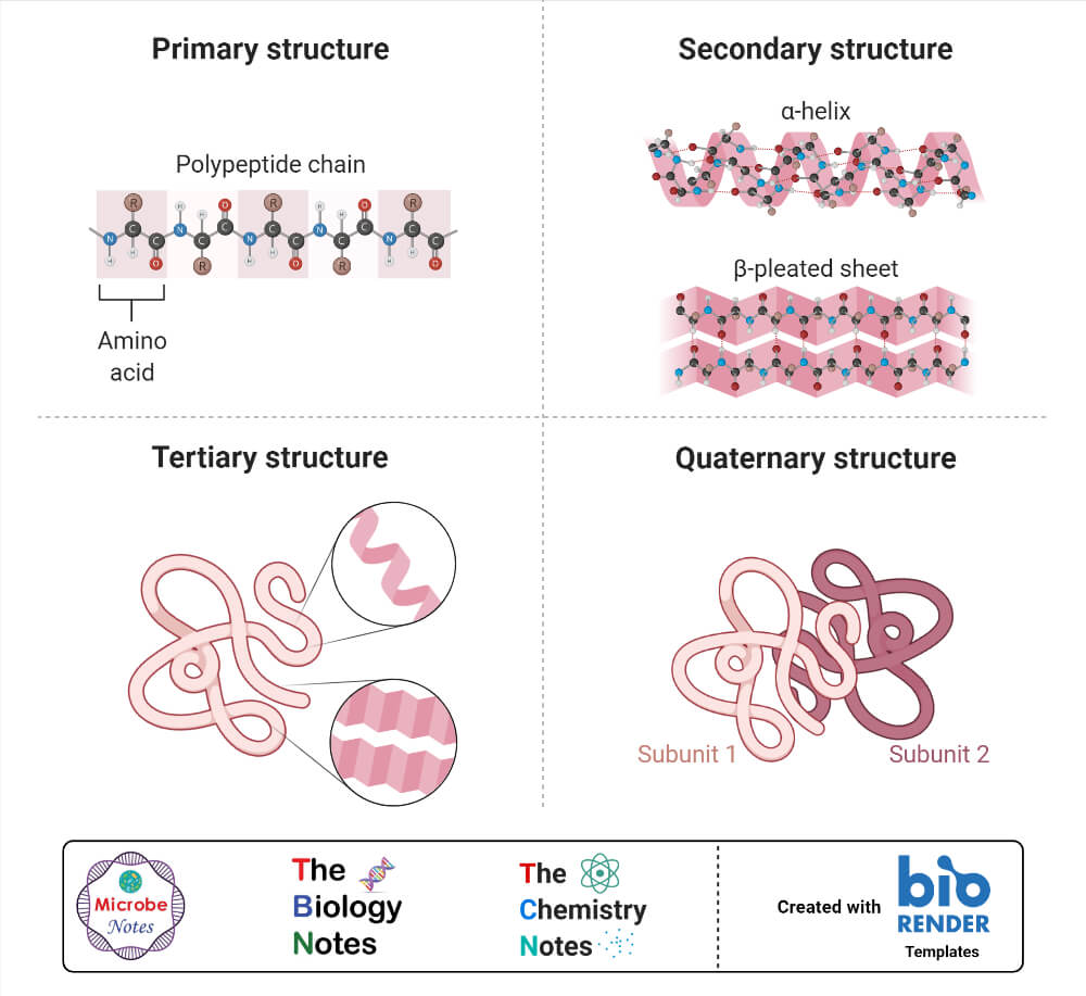
To understand how a protein achieves its final, functional shape, biologists describe its structure at four hierarchical levels: primary, secondary, tertiary, and quaternary. Let's explore each one.
1. Primary Structure: The Amino Acid Sequence
The journey of protein folding starts with its most fundamental level. The primary structure is simply the unique, linear sequence of amino acids in a polypeptide chain.
As we learned in the previous lesson, this sequence is determined by the genetic information encoded in a gene's DNA. This order is not random; it is the blueprint that dictates all subsequent levels of folding.
To see just how critical the primary structure is, let's examine a famous example: sickle cell anemia.
Protein structure: Primary, secondary, tertiary & quatrenary ...
This article from Khan Academy, 'Protein structure,' will be our main guide for this lesson. We'll start with the section on primary structure, which uses the excellent example of sickle cell anemia.
Read the section titled 'Primary structure'. Pay close attention to how a single amino acid change in the hemoglobin protein leads to sickle cell anemia.
As the article explains, changing just one amino acid (glutamic acid to valine) out of about 150 in the hemoglobin β chain is enough to cause the entire protein to fold incorrectly under certain conditions. This single error in the primary structure leads to malformed red blood cells and the serious health consequences of the disease. This powerfully illustrates that the primary structure is the absolute foundation upon which protein function is built.
2. Secondary Structure: Local Folds and Coils
Once the polypeptide chain is synthesized, it doesn't remain a straight line. It immediately begins to form local, repeating folded structures. This is the secondary structure.
These shapes are not determined by the R groups, but by hydrogen bonds forming between atoms of the polypeptide backbone—specifically, between the partially negative oxygen of a carbonyl group and the partially positive hydrogen of an amino group from another part of the chain.
The two most common types of secondary structure are:
- The α-helix: A delicate coil held together by hydrogen bonds between every fourth amino acid.
- The β-pleated sheet: Two or more segments of the polypeptide chain lying side-by-side, connected by hydrogen bonds.
Let's watch a video that visualizes the formation of these structures beautifully.
This video from Professor Dave Explains provides clear animations of how the α-helix and β-pleated sheet are formed and stabilized by hydrogen bonds along the polypeptide backbone.
Watch from 03:23 to 06:05. Focus on how the polypeptide backbone folds to form these two distinct shapes and where the hydrogen bonds form in each case.
Now, read the corresponding section in our main article for more detail, including how certain amino acids are more likely to be found in one structure versus the other.
Protein structure: Primary, secondary, tertiary & quatrenary ...
This section of the Khan Academy article provides further details on α-helices and β-pleated sheets.
Read the section titled 'Secondary structure'. Note the description of parallel and antiparallel β-sheets and the interesting role of proline as a 'helix breaker'.
3. Tertiary Structure: The Overall 3D Shape
While secondary structure describes local folding, tertiary structure refers to the overall, three-dimensional shape of a single polypeptide chain. This is where the protein starts to look like a complex, functional object.
This level of folding is driven primarily by interactions between the R groups (side chains) of the various amino acids. Several types of interactions work together to stabilize the tertiary structure:
- Hydrophobic Interactions: Amino acids with nonpolar, hydrophobic R groups tend to get pushed to the interior of the protein to hide from the surrounding water. This is a major driving force in protein folding.
- Hydrogen Bonds: Form between polar R groups.
- Ionic Bonds: Form between positively and negatively charged R groups (acidic and basic side chains).
- Disulfide Bridges: A strong, covalent bond that can form between the side chains of two cysteine amino acids. These act like "molecular safety pins," reinforcing the protein's shape.
Protein structure: Primary, secondary, tertiary & quatrenary ...
Now let's read about the forces that shape the final fold of a polypeptide.
Read the section 'Tertiary structure'. The diagram in this section is particularly helpful for visualizing the different types of R-group interactions.
4. Quaternary Structure: Assembling Multiple Chains
Some proteins consist of a single polypeptide chain, and for them, the tertiary structure is the final level. However, many proteins are made of two or more polypeptide chains, called subunits.
The quaternary structure describes how these subunits are arranged and interact to form the final, functional protein complex. The same types of interactions that stabilize tertiary structure (hydrogen bonds, ionic bonds, etc.) also hold these subunits together.
The classic example is hemoglobin, which we discussed earlier. It is composed of four subunits: two α-globin and two β-globin chains. Only when all four are assembled correctly can it effectively transport oxygen.
Protein structure: Primary, secondary, tertiary & quatrenary ...
Finally, let's look at the highest level of protein structure.
Read the section titled 'Quaternary structure'. Notice that the same forces that create tertiary structure are also at play here.
This image provides a great visual summary of all four levels of protein structure we've discussed.
Test your understanding!
For each statement below, identify which level(s) of protein structure (Primary, Secondary, Tertiary, Quaternary) it describes.
- Stabilized by hydrogen bonds between atoms of the polypeptide backbone.
- The sequence of amino acids in a protein.
- A multimeric protein like hemoglobin, made of four polypeptide subunits.
- Determined by interactions between the R groups of amino acids, including disulfide bridges.
- A change at this level can affect all other levels.
Show answer
- Secondary structure. This is the key distinction from tertiary structure, which involves R-group interactions.
- Primary structure. This is the fundamental blueprint.
- Quaternary structure. This level specifically describes proteins with multiple subunits.
- Tertiary structure (and also helps stabilize Quaternary structure). These R-group interactions create the overall 3D fold.
- Primary structure. A change in the amino acid sequence can alter folding at every subsequent level.
Structure Determines Function
We've now seen how a protein folds. This final, specific 3D shape, or conformation, is what allows a protein to do its job. An enzyme has an active site with a specific shape to bind its substrate. An antibody has a binding site shaped to recognize a specific antigen. The relationship is direct: Sequence → Structure → Function.

What happens if this delicate structure is disrupted? This is called denaturation. Exposure to non-optimal conditions, like high temperatures, extreme pH, or certain chemicals, can break the weak interactions (hydrogen and ionic bonds) that hold a protein's shape. The protein unravels, losing its secondary, tertiary, and quaternary structure. A denatured protein is non-functional.
A great real-world example is frying an egg. The clear egg white is mostly water and a protein called albumin. The heat from the pan denatures the albumin, causing it to unfold and tangle together, forming the solid, opaque white we see.
Protein structure: Primary, secondary, tertiary & quatrenary ...
Let's conclude by reading about denaturation.
Read the section 'Denaturation and protein folding'. Notice the distinction between reversible and irreversible denaturation.
Conclusion
Today, we journeyed from a simple polypeptide chain to a complex, functional protein. We saw how a precise hierarchy of folding events, governed by chemical principles, creates the amazing diversity of shapes and functions found in the protein world.
Key Takeaways:
- Primary structure is the linear sequence of amino acids.
- Secondary structure consists of local α-helices and β-pleated sheets, stabilized by backbone hydrogen bonds.
- Tertiary structure is the overall 3D shape of a single polypeptide, determined by R-group interactions (hydrophobic interactions, hydrogen bonds, ionic bonds, disulfide bridges).
- Quaternary structure is the assembly of multiple polypeptide subunits into a larger protein complex.
- A protein's specific 3D conformation is essential for its function.
- Denaturation is the loss of this structure and function due to environmental stress.
Preview of the next lesson:
We have now completed our tour of the four major classes of biological macromolecules. With this foundational knowledge of the cell's chemical components, we are ready to explore the cell itself. In the next module, we will begin our "Tour of the Cell," starting with the fundamental differences between prokaryotic and eukaryotic cells.