Hello! Welcome back to our course.
In our last lesson, we learned how to interpret AI-generated differential diagnoses for oral lesions, looking at everything from probability scores to the importance of explainability tools like heatmaps. We established that AI can be a powerful assistant in the diagnostic process.
Today, we'll narrow our focus to one of the most critical applications of this technology: oral cancer screening. The goal of this lesson is to describe AI applications in oral cancer screening using clinical photographs. We will explore how AI analyzes these images, see real-world examples of how this is deployed (especially via smartphones), and understand how it functions as a clinical decision support tool. With oral cancer rates projected to rise globally, early and accurate detection is more vital than ever, and AI is emerging as a key ally.
1. Traditional vs. AI-Powered Screening
The cornerstone of oral cancer detection has always been the Clinical Oral Examination (COE), which relies on a clinician's ability to visually inspect and palpate tissues. While indispensable, its effectiveness can vary based on experience and lighting conditions. AI introduces a new layer of objectivity and consistency.
To get a clear overview of how AI-powered screening compares to the traditional approach, let's start with a resource that breaks down the key differences.
AI vs. Traditional Oral Cancer Screening - Complete Smiles
The article 'AI vs. Traditional Oral Cancer Screening' from Complete Smiles provides a concise comparison and introduces the fundamental concepts of AI-based screening.
First, read the introductory section, paying close attention to the 'Quick Comparison' table. Then, read the section 'What Is AI-Based Oral Cancer Screening?' to understand the underlying technology. Finally, review the first question in the 'FAQs' section, which directly summarizes how AI enhances screening.
As the article explains, AI leverages machine learning—specifically, a type of neural network called a Convolutional Neural Network (CNN) that we've encountered before—to analyze images. These models are trained on vast datasets containing thousands of clinical photographs of benign, precancerous, and cancerous lesions. By processing these examples, the AI learns to identify the subtle patterns, textures, and color variations that are often invisible to the naked eye but may indicate malignancy.
The process begins with acquiring a high-quality clinical photograph, a step that has become incredibly accessible.

Once the image is captured, the AI model processes it to classify the lesion, acting as an objective "second pair of eyes."
2. Real-World Applications: Screening in Your Pocket
The true power of using AI for oral cancer screening lies in its accessibility. By developing AI models that can run on smartphones, this diagnostic support can be deployed far beyond the specialist's office.
This approach is particularly promising for improving healthcare equity, as confirmed by a recent review highlighted by the American Dental Association (ADA).
AI shows promise in early detection of oral cancer review finds
This ADA News article summarizes a scoping review on AI in oral cancer detection, confirming the potential of using mobile images.
Please read the second and third paragraphs of the article. Focus on the 'common data sources' mentioned and the role of smartphone apps in low-resource settings.
The ADA article reinforces that clinical photographs and mobile images are primary data sources for these AI systems. This has led to the development of powerful real-world applications.
Let's look at a specific example of such a project.
AI vs. Traditional Oral Cancer Screening - Complete Smiles
Returning to the 'Complete Smiles' article, let's examine a concrete example of a mobile screening initiative.
Read the section 'Applications in Clinical Settings'. Pay close attention to the description of the 'Mobile Mouth Screening Anywhere (MeMoSA)' initiative.
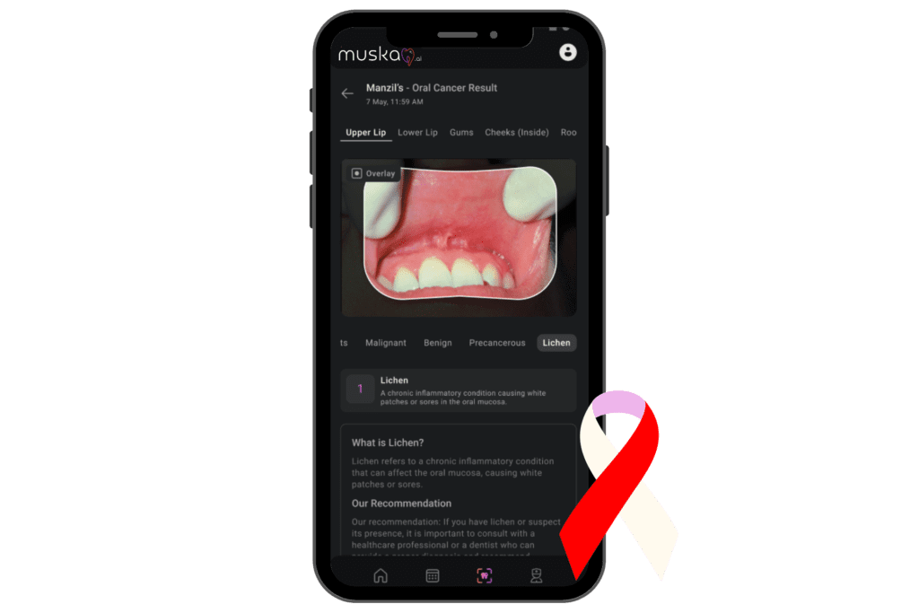
The MeMoSA project is a perfect illustration of this lesson's core concept. It uses a deep learning model to classify oral lesions from smartphone images, with the specific goal of improving screening access in underserved communities. A health worker with a smartphone can capture an image of a suspicious lesion and get an immediate, AI-driven risk assessment.
The output from such an app provides a preliminary classification and guides the user on the next steps.
3. Building Trust: From Black Box to Diagnostic Assistant
A major hurdle for clinical adoption is the "black box" problem: how can a clinician trust a recommendation without knowing the AI's reasoning? This is where Explainable AI (XAI) becomes crucial, a concept we touched on in our last lesson.
In the context of oral cancer screening, XAI provides visual evidence for the AI's findings.
AI vs. Traditional Oral Cancer Screening - Complete Smiles
Let's read one final section that discusses the future of these tools and the importance of explainability.
Read the section 'Future Role of AI in Oral Precancerous Detection', focusing on the final paragraph about 'Explainable AI (XAI)' and the use of heatmaps.
As the article mentions, tools like heatmaps (or saliency maps) can be overlaid on the original clinical photograph to highlight the specific pixels or regions that the AI found most suspicious. If the AI flags a lesion as potentially malignant, the heatmap would show exactly which part of the lesion's morphology (e.g., an area of ulceration or an unusual color patch) contributed most to that decision.
This transparency achieves two things:
- Builds Trust: It allows you, the clinician, to validate the AI's "thinking" against your own clinical knowledge.
- Enhances Learning: It can draw your attention to subtle features you may have overlooked, improving your own diagnostic acumen over time.
Ultimately, the goal is not to replace the clinician but to augment their abilities. The AI serves as a tireless, objective screening tool that can flag at-risk patients early, especially in settings where a specialist may not be immediately available.
Conclusion
In this lesson, we've seen how AI is being applied to one of the most important screening tasks in dentistry. By analyzing simple clinical photographs, these systems offer a scalable, accessible, and objective method for the early detection of oral cancer.
Key Takeaways:
- Core Technology: AI uses Convolutional Neural Networks (CNNs) trained on thousands of labeled clinical photographs to identify visual patterns associated with oral potentially malignant disorders and cancer.
- Primary Application: The most significant application is through mobile health (mHealth) platforms. Smartphone apps can empower general dentists, hygienists, and community health workers to perform initial screenings and triage cases effectively.
- Accessibility and Equity: This technology holds immense promise for improving early detection rates in remote or underserved areas where access to specialists is limited.
- Clinical Role: AI functions as a decision support tool, not a replacement for clinical judgment. Its role is to act as an objective "second opinion."
- Building Trust: Explainable AI (XAI) features, such as heatmaps, are essential for clinical adoption as they make the AI's reasoning transparent and verifiable.
Preview of the Next Lesson
Having concluded our module on AI in advanced diagnostics, we will now shift our focus from diagnosis to treatment. In the first lesson of our next module, "AI-Assisted Treatment Planning," we will explore how AI automates cephalometric landmark identification—a foundational task in orthodontics that saves clinicians time and improves consistency.