Hello! Welcome back.
In our previous lesson, we explored how AI assists in detecting and characterizing periapical lesions. We focused on a relatively defined task: identifying a specific type of lesion. Now, we will broaden our scope to a more complex and common clinical challenge.
When you encounter an unusual lesion in a patient's mouth, your clinical reasoning kicks in to form a differential diagnosis—a list of possible conditions that could explain the finding. Today's lesson is about how AI can act as a sophisticated assistant in this process. Our learning outcome is to interpret AI-generated outputs that suggest a differential diagnosis for oral lesions. We will look at two main types of AI systems: computer vision models that analyze images and large language models that process text-based clinical descriptions.
1. The Diagnostic Challenge of Oral Lesions
Forming a differential diagnosis for oral lesions is one of the most challenging aspects of clinical dentistry. Many different conditions—from harmless irritations to life-threatening cancers—can appear visually similar, especially in their early stages.
To appreciate the complexity that AI aims to help with, let's start with a review article that sets the scene.
Artificial intelligence and the diagnosis of oral cavity cancer and oral ...
The following paper, 'Artificial intelligence and the diagnosis of oral cavity cancer and oral potentially malignant disorders from clinical photographs', provides an excellent overview of the diagnostic challenge.
Please read the first three paragraphs of the 'Introduction'. Focus on the descriptions of the clinical heterogeneity of oral potentially malignant disorders (OPMDs) and the difficulties non-specialists face in triaging lesions.
As the paper highlights, the sheer variety in how these lesions present makes it difficult to distinguish between benign, premalignant, and malignant conditions based on visual inspection alone. This is where AI can offer powerful support. Instead of just performing a binary task (e.g., "lesion present/absent"), these advanced AI models perform multi-class classification. They are trained not just to see a lesion, but to predict which of several possible diagnostic categories it belongs to.
2. Interpreting Outputs from Computer Vision Models
The most common way AI assists with differential diagnosis is by analyzing clinical photographs or radiographs. Let's break down what their outputs look like and, more importantly, how to interpret them with a critical clinical eye.
What Do the Outputs Look Like?
An AI model's "suggestion" for a differential diagnosis typically comes in one of a few formats:
- A Categorized List: The AI classifies the lesion into a predefined category.
- A Ranked List with Probabilities: The AI provides a list of possible diagnoses, each with a corresponding probability score.
Here is an example of what a user interface for such an AI might look like on a smartphone.
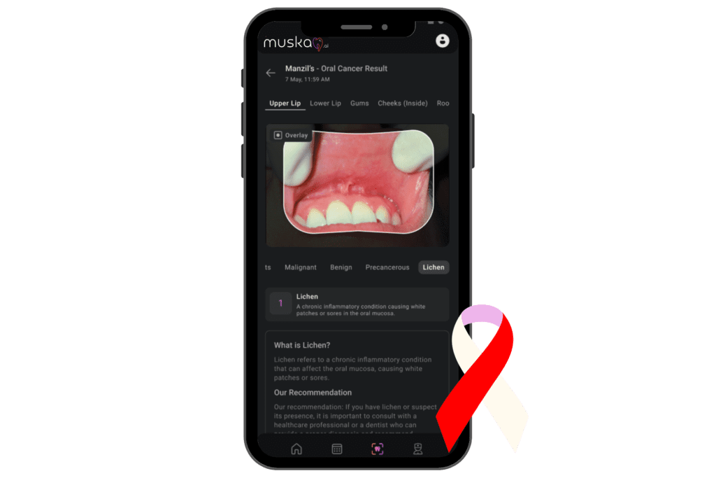
This output is a starting point for your differential diagnosis. The AI is suggesting "Lichen" but also implicitly offering the other categories as possibilities. A more detailed output would rank these possibilities with scores, for example:
- Lichen Planus: 75%
- Precancerous (Leukoplakia): 15%
- Benign Frictional Keratosis: 8%
- Malignant (OSCC): 2%
How to Critically Interpret the Outputs
A list of probabilities can be powerful, but it can also be misleading if not interpreted correctly. Simply trusting the top result is not enough. You need to understand why and how confidently the AI made its suggestion.
Artificial intelligence and the diagnosis of oral cavity cancer and oral ...
To understand how to look 'under the hood' of an AI's prediction, let's return to the review paper on AI for oral cancer diagnosis. It discusses the critical concepts of explainability and uncertainty.
Please read the section '3.7 Explainability, uncertainty, and transparency'. Pay close attention to the descriptions of saliency maps (like Grad-CAM) and uncertainty quantification. Then, read the first three paragraphs of 'Section 3: discussion' (which is section 4 of the article), which highlight the clinical need for multi-class systems over simple binary classifiers.
Based on this reading, let's establish a framework for interpreting these outputs:
1. Scrutinize the Probability Score:
As the paper notes, a probability score is not the same as the model's confidence. A model might assign an 80% probability to a diagnosis simply because it's the "least bad" option among the classes it knows, even if the model is very uncertain overall.
2. Demand an Explanation (Explainability):
How can you trust the AI's reasoning? You need to see its evidence.
- Saliency Maps (Heatmaps): These are one of the most important explainability tools. A heatmap visually overlays the input image, highlighting the pixels the AI considered most important for its decision.
- Your Action: If the AI suggests "OSCC" but the heatmap is focused on a dental restoration or a tongue papilla instead of the lesion itself, you should be highly skeptical of its suggestion. The AI may have been distracted by an irrelevant feature.
3. Ask "How Sure Are You?" (Uncertainty Quantification):
Some advanced models can provide a separate uncertainty score. This is more direct than trying to infer confidence from probability.
- Low Uncertainty: The model is confident in its prediction. The features it saw strongly match what it learned during training for that specific diagnosis.
- High Uncertainty: The model is "confused." The lesion may have ambiguous features, look like multiple conditions at once, or be something the AI has never seen before.
- Your Action: A high uncertainty score is a red flag. It's a direct message from the AI to you, the clinician, that this case requires extra scrutiny, further investigation, and perhaps referral.
The paper's discussion reinforces a key clinical point: the utility of an AI tool depends entirely on whether it's trained to answer a meaningful question. A simple "cancer vs. normal" classifier is far less useful than a multi-class system that can differentiate between OSCC, OPMDs, and common benign look-alikes, which is the essence of building a differential diagnosis.
3. Interpreting Outputs from Large Language Models (LLMs)
A different class of AI, Large Language Models (LLMs) like ChatGPT, can also generate differential diagnoses. Instead of analyzing images, they analyze textual information—your written description of a lesion, along with the patient's history.
ChatGpt's accuracy in the diagnosis of oral lesions - PMC
A recent study evaluated how accurately ChatGPT can diagnose oral lesions based on clinical scenarios. Let's look at their findings.
First, read the 'Introduction' to understand how LLMs can fit into the diagnostic workflow. Then, review the 'Results' section to see the accuracy rates. Finally, read the 'Discussion', paying close attention to the acknowledged limitations, such as the inability to perform a physical exam and the lack of visual data.
From this study, we can establish a similar interpretation framework for LLMs:
1. Acknowledge the Accuracy Limits:
The study found an overall accuracy of 87.4% for ChatGPT-4.0, which dropped to 78.4% for difficult questions. This is a crucial takeaway: LLMs are not infallible. Their suggestions must be verified.
2. Address the "Hallucination" Problem:
LLMs can sometimes generate plausible but incorrect information, a phenomenon known as "hallucination." How can we ensure the information is grounded in medical fact? This is where a technology called Retrieval-Augmented Generation (RAG) comes in.
Artificial intelligence in digital pathology
To understand how to make an LLM more trustworthy, let's watch a short clip from the video 'Artificial intelligence in digital pathology'. It explains the concept of RAG in a very clear, conceptual way.
Watch the segment from 05:21 to 06:11. It describes how RAG forces an LLM to base its answers on a solid database and to provide references for its claims.
RAG transforms an LLM from a creative storyteller into a research assistant. By forcing the model to cite its sources from a trusted database (e.g., medical literature), it dramatically increases the reliability of its diagnostic suggestions. When interpreting an LLM's output, look for citations or references that back up its claims.
3. Respect the Inherent Limitations:
The research paper on ChatGPT is very clear about the model's limitations, which are fundamental to your interpretation:
- No Physical Exam: An LLM cannot palpate a lesion to assess its texture or firmness.
- No Visual Data (Usually): Unless it's a multimodal model (like GPT-4V), it cannot see the lesion's color, shape, or surface characteristics.
- No Follow-up Questions: The AI cannot ask for clarification as a human clinician would.
Your Action: The output from an LLM is based only on the text you provide. If your description is incomplete or ambiguous, the AI's suggestions will be flawed. The responsibility for providing high-quality input rests entirely on you.
Conclusion
AI is emerging as a valuable tool for building a differential diagnosis for oral lesions. It can process vast amounts of data to find patterns and suggest possibilities that might be missed, especially by a non-specialist.
Key Takeaways:
- How AI Suggests a Differential Diagnosis:
- Computer Vision Models: Use multi-class classification on images to produce a ranked list of diagnoses with probability scores.
- Large Language Models: Analyze text-based clinical scenarios to generate a list of possible diagnoses.
- How to Interpret the Outputs:
- Look beyond the top suggestion. Critically assess probability scores, but do not equate them with confidence.
- Use explainability tools like saliency maps to verify the AI's visual reasoning.
- Pay attention to uncertainty scores as a direct measure of the model's confidence.
- For LLMs, verify claims using technologies like RAG that ground answers in reliable sources.
- The Clinician is the Final Authority: AI outputs are powerful suggestions, but they are not final diagnoses. They must be integrated with your own clinical examination, patient history, and professional judgment.
Preview of the Next Lesson
We've now seen how AI can help formulate a differential diagnosis. In our next lesson, we will focus on one of the most critical applications of this technology: AI applications in oral cancer screening using clinical photographs. We will delve into how these tools are being deployed specifically to tackle the challenge of early cancer detection.