Hello! Welcome to the first lesson in our new module, "AI in Advanced Diagnostics and Pathology."
In our last lesson, we concluded our exploration of AI for caries and periodontal disease by focusing on the common limitations of these systems, such as detecting early lesions and handling imaging artifacts. This critical perspective is essential as we move into more complex diagnostic tasks.
Today, we'll apply what we've learned to the field of endodontics. Our learning outcome is to explain how AI assists in detecting and characterizing periapical lesions on radiographs and CBCT. A periapical lesion (PAL) is an area of inflammation around the apex of a tooth's root, typically resulting from a pulpal infection. Detecting these lesions accurately is crucial for diagnosis, treatment planning, and evaluating the success of endodontic therapy. We will explore how AI tackles this challenge in both 2D and 3D imaging.
1. The Challenge of Diagnosing Periapical Lesions
Before we look at AI's role, it's important to understand why detecting PALs can be difficult for human clinicians.
Detection of Periapical Lesions Using Artificial Intelligence - PMC
To understand the clinical problem that AI aims to solve, let's start with a section from a comprehensive review paper titled 'Detection of Periapical Lesions Using Artificial Intelligence'.
Please read the first and second paragraphs of the 'Introduction'. These paragraphs define periapical lesions (PALs) and describe the pros and cons of the different imaging methods used to detect them (IOPA, OPG, and CBCT).
As the paper explains, traditional 2D radiographs (IOPAs and OPGs) suffer from anatomical superimposition, which can obscure lesions. While 3D CBCT scans are far more accurate, they involve a higher radiation dose and cost, and interpreting the large volume of data can be time-consuming and subject to observer variability.
These challenges create a perfect opportunity for AI to act as an assistive tool, helping to improve both the accuracy and efficiency of diagnosis.
2. How AI Assists with 2D Radiographs (IOPA & OPG)
At a high level, an AI model analyzes a dental radiograph through a series of steps to identify a potential lesion. This process is conceptually similar to what we've discussed for caries detection.
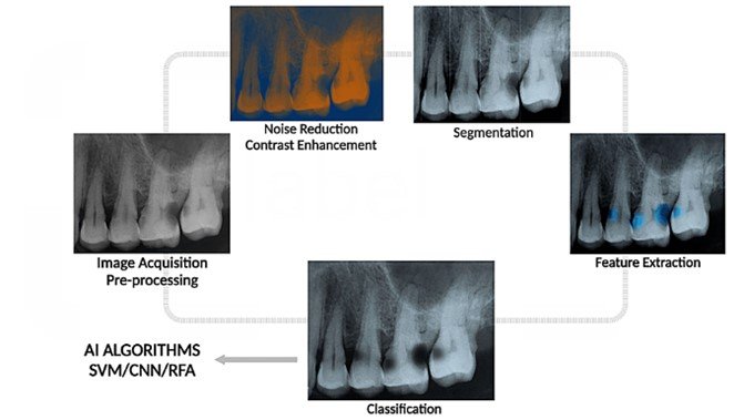
Let's see how this is applied to the two main types of 2D radiographs.
Panoramic Radiographs (OPGs)
OPGs provide a broad overview of the entire mouth, making them excellent for initial screening. AI models can analyze these large images very quickly.
Detection of Periapical Lesions Using Artificial Intelligence - PMC
The same review paper we just looked at also details AI's performance on panoramic images. Let's read the relevant section.
Please read section '3.1. AI-Based Systems Performance for Detecting Periapical Lesions Using Panoramic Images (OPG)'. Focus on the key findings regarding AI's speed and its performance compared to clinicians.
The key takeaways for OPGs are:
- Speed: AI can analyze a panoramic image in seconds, whereas a human might take several minutes.
- Performance: Studies show AI systems can outperform less experienced dentists and serve as a reliable screening tool.
- Limitation: Similar to humans, AI struggles with the inherent low resolution and superimposition of OPGs. Its accuracy on OPGs is lower than on CBCTs.
Periapical Radiographs (IOPAs)
IOPAs are the standard for detailed endodontic assessment. AI models trained on these images can achieve very high accuracy in detecting PALs.
AI-Powered Clinical Decision Support Tools in Endodontic Education
To see a practical example and learn about AI's performance on IOPAs, let's look at an article from the American Association of Endodontists (AAE) and then return to our review paper.
First, read the introduction of this article up to and including Figure 1. The figure shows a real-world example of an AI (Overjet) highlighting a periapical radiolucency. Then, read the first paragraph of the main body, which discusses how AI tools are trained for this task and cites performance data.
The AAE article highlights that AI can act as a "failsafe" to catch lesions that might otherwise be missed. The review paper provides more detail on the specific models and their performance.
Detection of Periapical Lesions Using Artificial Intelligence - PMC
Now, let's examine the research findings on IOPA performance in more detail.
Please read section '3.2. AI-Based Systems Performance for Detecting Periapical Lesions Using Periapical Radiographs'. This section describes the various AI architectures used and their reported accuracies.
For IOPAs, AI demonstrates strong diagnostic performance, often using object-detection models like YOLO or segmentation models like U-Net to precisely outline the lesion.
3. Stepping into the Third Dimension: AI and CBCT
CBCT is considered the reference standard for diagnosing PALs due to its 3D nature, which eliminates anatomical overlap. However, interpreting a full CBCT volume slice by slice is time-consuming. This is where AI offers a significant advantage in efficiency.
Detection of Periapical Lesions Using Artificial Intelligence - PMC
Let's explore AI's role in analyzing these complex 3D scans.
Please read the section '3.3. AI-Based Systems Performance for Detecting Periapical Lesions Using CBCT Images'. Focus on why AI is a promising adjunct for CBCT analysis and the reported diagnostic accuracy.
AI systems for CBCT, often using 3D CNNs, analyze the entire volumetric dataset to detect and segment lesions. Studies show this assistance not only improves accuracy but also dramatically reduces interpretation time for both junior and senior clinicians.
4. Characterizing Lesions: The Clinical Nuances
Beyond simply detecting a lesion, AI can help characterize it. However, its performance is influenced by several clinical factors, echoing the limitations we discussed in the previous lesson.
Lesion Size
Just as AI struggles with incipient caries, it is less sensitive to small, early periapical lesions.
Detection of Periapical Lesions Using Artificial Intelligence - PMC
Let's look at how lesion size impacts AI performance.
Please read the section '5. Influence of Periapical Lesion Size on AI Performance'. Notice the significant drop in sensitivity for lesions below a certain size threshold.
This finding is critical for clinical practice: you cannot rely on AI to catch every subtle, early change in the periapical bone.
Anatomical Complexity
The location of the tooth also matters. Areas with complex or overlapping anatomy can confuse the AI.
Detection of Periapical Lesions Using Artificial Intelligence - PMC
The anatomical neighborhood of a tooth root can pose a significant challenge for AI.
Please read the section '6. Influence of Tooth Type and Anatomical Surroundings on AI Performance'. Pay close attention to the example of the maxillary sinus, a common source of diagnostic confusion.
The proximity of the maxillary sinus floor to the root apices of upper molars can mimic a lesion, leading to false positives or negatives for both humans and AI. This highlights the need for careful clinical correlation.
Root-Filled Teeth and Artifacts
The presence of a root canal filling introduces another layer of complexity, particularly on CBCT scans.

Detection of Periapical Lesions Using Artificial Intelligence - PMC
The presence of root filling material has a dual effect on AI performance.
Please read the section titled '4.1. Influence of Root-Filling Status on AI Performance'. This section explains how fillings can sometimes help the AI by providing a clearer border, but can also hurt performance by creating artifacts.
This is a perfect example of a clinical trade-off. While the radiopaque gutta-percha can enhance the contrast between the root and a lesion, the metallic artifacts it may cause can lead to diagnostic errors.
Conclusion
AI is rapidly becoming a powerful co-pilot for detecting and characterizing periapical lesions. It can process images with incredible speed, act as a failsafe to prevent missed diagnoses, and improve the accuracy of clinicians at all experience levels.
Key Takeaways:
- Detection Across Modalities: AI assists in PAL detection by analyzing 2D radiographs (OPG, IOPA) and 3D CBCT scans to identify and outline areas of periapical radiolucency.
- Characterization Limitations: AI's ability to characterize lesions is influenced by clinical factors. Its performance is higher for larger lesions and can be reduced by anatomical complexity (like the maxillary sinus) and artifacts from root fillings.
- AI as an Augmentation Tool: AI is not an autonomous diagnostician. It is a decision-support tool that augments the clinician's abilities. Its findings must always be integrated with clinical examination, patient history, and professional judgment. As the AAE article warns, you must treat the patient, not the radiograph.
Preview of the Next Lesson
We've seen how AI can detect and outline a specific pathology like a periapical lesion. But what happens when the radiographic signs are less clear? In our next lesson, we will explore our next learning outcome: Interpret AI-generated outputs that suggest a differential diagnosis for oral lesions. We'll move beyond simple detection to see how AI can help clinicians consider a range of possible diagnoses for a given finding.