Hello! Welcome to the first lesson in our new module on AI-Assisted Treatment Planning.
In our previous module, we concluded by examining the significant challenges AI faces in high-stakes diagnostic areas like oral pathology, including data scarcity, bias, and the "black box" problem. We noted that AI's path to adoption isn't always straightforward.
Today, we'll explore a different, highly successful application of AI that provides a fascinating contrast. Instead of making a complex diagnosis, this AI task is focused on automating a routine, time-consuming, but critical step in orthodontic planning. Our goal for this lesson is to describe how AI automates cephalometric landmark identification for orthodontics. You will learn what cephalometric analysis is, why the manual process is challenging, and how AI has transformed it into a task that can be done in seconds.
1. What is Cephalometric Analysis?
Before we can understand how AI helps, we must first understand the clinical task it's designed to perform. Cephalometric analysis is a cornerstone of orthodontic diagnosis and treatment planning. It involves taking a standardized x-ray of the head (a cephalogram) and identifying specific anatomical points, or "landmarks."
To get a practical understanding of these landmarks and their importance, let's watch a video from a familiar dental education channel.
Orthodontics | Diagnosis & Treatment Planning | INBDE, ADAT
This video from the 'Mental Dental' YouTube channel offers an excellent clinical overview of cephalometrics. It details the key landmarks and explains why orthodontists use them.
Please watch the segment from 00:31:59 to 00:42:38. The first part (00:31:59 - 00:38:09) identifies numerous anatomical landmarks. You don't need to memorize every single one, but focus on understanding what they represent and recognizing key points like Sella (S), Nasion (N), A point, and B point. The second part (00:38:58 - 00:42:38) explains how these points create reference planes and angles (like SNA, SNB, and ANB) that are used for diagnosis.
As the video explains, once these landmarks are identified, they are connected to form lines and planes. The angles and distances between them allow an orthodontist to quantitatively assess a patient's skeletal jaw relationships, tooth positions, and soft tissue profile. This analysis is fundamental for diagnosing conditions like a prognathic mandible (Class III) or retrognathic mandible (Class II) and for planning treatments, including orthodontics or orthognathic surgery.
2. The Challenge of Manual Tracing
The traditional process of cephalometric analysis involves an orthodontist manually identifying each of these landmarks. Historically, this was done by tracing on acetate paper placed over the x-ray. More recently, it's done by clicking on a digital image. While this is a standard procedure, it has several well-known drawbacks.
Let's read the introduction to a recent scientific review that perfectly frames the problem.
[PDF] Deep Learning in Cephalometric Analysis: A Scoping Review of ...
This paper, 'Deep Learning in Cephalometric Analysis', starts by explaining the core challenges of the manual method, which motivated the development of automated solutions.
Please read the 'Abstract' and 'Introduction' sections of the paper. Focus on the descriptions of manual tracing as a 'labor-intensive process prone to inter-observer variability and human error'.
The reading highlights the three main challenges of manual landmarking:
- It's time-consuming: Manually locating 20, 30, or even more points on a single radiograph is a tedious task that takes up valuable clinical time.
- It requires significant expertise: Accurately and consistently identifying these landmarks, especially in radiographs with poor image quality or overlapping structures, requires years of training and experience.
- It's subjective and prone to error:
- Inter-observer variability: Two different expert orthodontists may place the same landmark in slightly different positions.
- Intra-observer variability: A single orthodontist might place a landmark differently if they trace the same radiograph on two separate occasions.
These inconsistencies, while often small, can affect the resulting measurements and potentially influence treatment planning. This combination of being slow, difficult, and subjective makes cephalometric tracing a prime candidate for AI automation.
3. The AI Solution: Automated Identification
AI, specifically deep learning, provides a powerful solution to these challenges. The goal is to train a model that can look at a cephalometric radiograph and automatically output the precise coordinates of all the required landmarks.
How It Works: A Conceptual View
At a high level, the process is similar to how we've seen AI work on other image-based tasks. The core technology used is typically a Convolutional Neural Network (CNN).
Let's look at a diagram that breaks down the process.
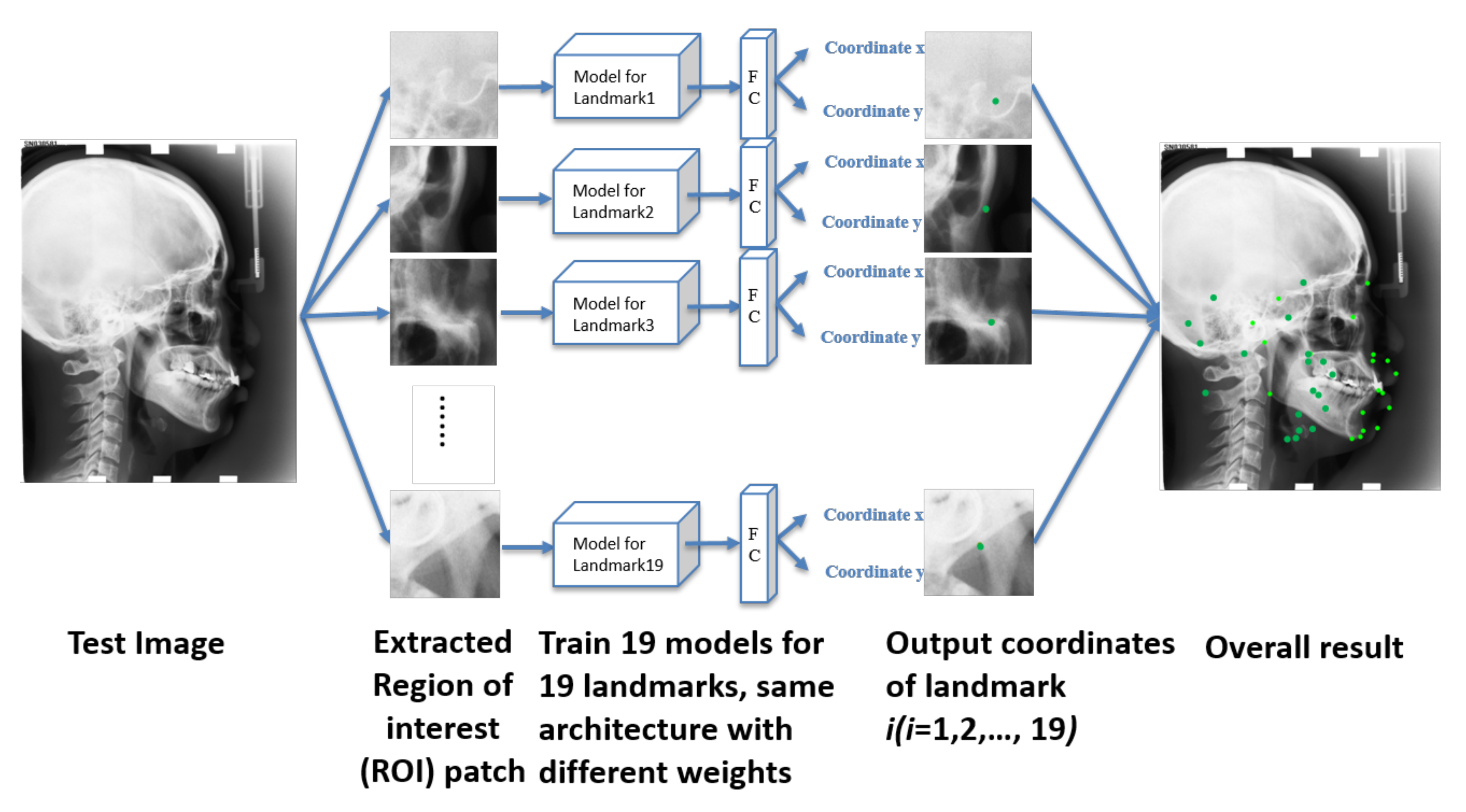
Here’s a step-by-step breakdown of how this is achieved:
- Training Data: The AI model is trained on a massive dataset, often thousands of cephalograms. For each image in this dataset, one or more expert orthodontists have already manually marked the correct location of every landmark. This expert-labeled data serves as the "ground truth."
- Learning Patterns: The CNN learns the complex visual patterns associated with each landmark. For instance, it learns to recognize the specific saddle-like shape of the sella turcica to find the "Sella" point and the junction between the frontal and nasal bones to find the "Nasion" point.
- Prediction: Once trained, the AI can be given a new, unseen cephalogram. It applies what it has learned to predict the (x, y) coordinates for each landmark. The entire process takes only a few seconds.
The result is a fast, objective, and consistent analysis. The image below shows a direct comparison between landmarks identified by a human expert and those identified by an AI.
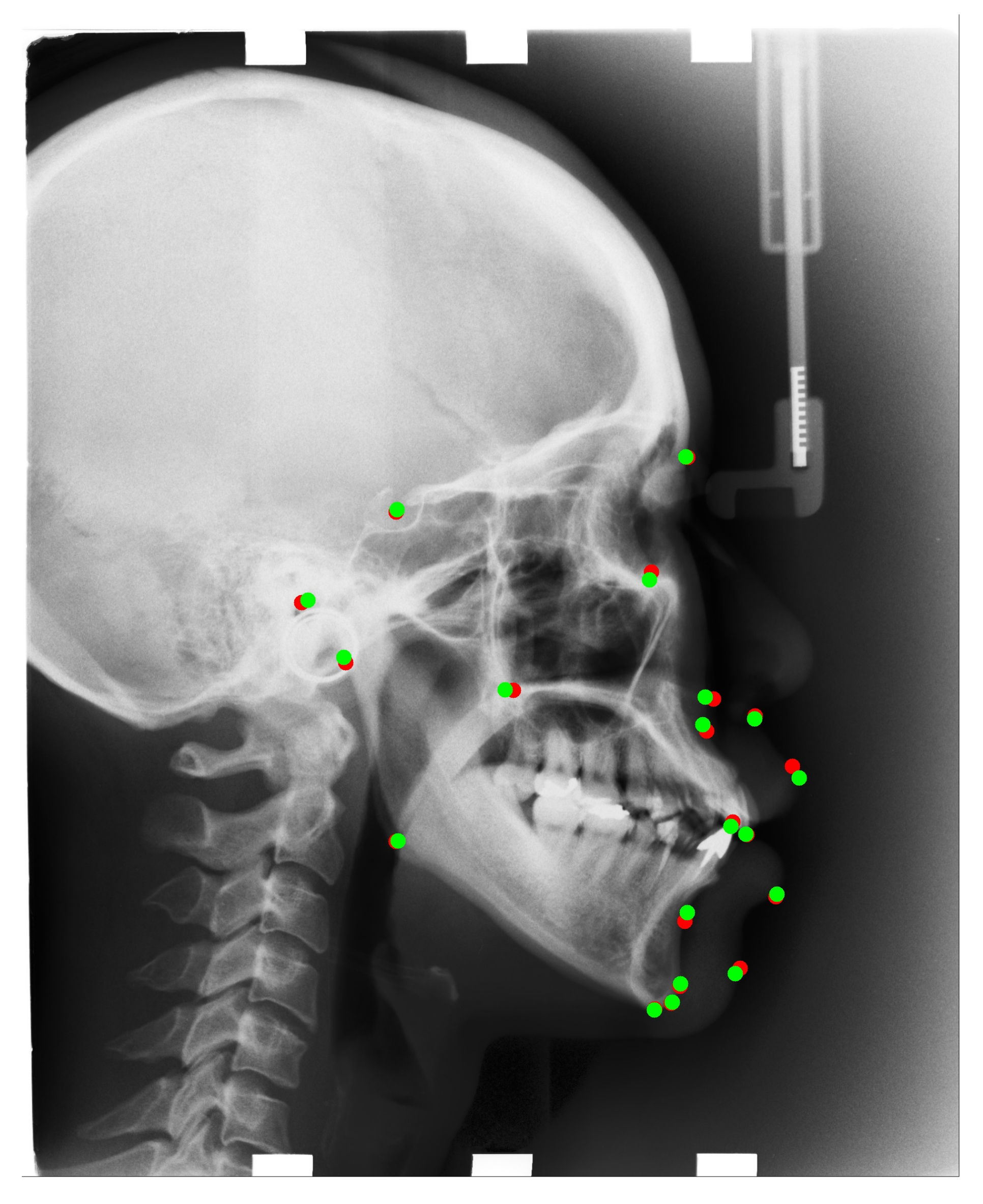
The technology to do this has improved dramatically. Let's briefly look at the evolution of these methods.
[PDF] Deep Learning in Cephalometric Analysis: A Scoping Review of ...
The technology behind this automation has evolved rapidly. This section from the same review paper gives a concise history of the methods used.
Please read the section titled 'B. Landmark Detection Evolution'. You don't need to understand the technical jargon for older methods like 'ASMs' or 'HOG,' but focus on the main point: since 2015, Deep Learning and CNNs have become the dominant and most accurate approach.
As the paper notes, this technology is no longer just in research labs. Numerous commercial software products, such as WebCeph, CephX, AudaxCeph, and others, now offer AI-powered automated cephalometric analysis to clinicians worldwide.
4. Benefits of AI Automation
The impact of this technology on orthodontic workflow is significant. Let's summarize the key benefits:
- Efficiency: The most obvious benefit is speed. A task that takes a clinician several minutes can be completed by an AI in under a second. A study by Park et al. (2019), discussed in the review "Application of Artificial Intelligence (AI) in a Cephalometric Analysis," found that the YOLOv3 algorithm could perform the analysis in just 0.05 seconds.
- Consistency: AI removes the subjectivity inherent in manual tracing. An AI model will produce the exact same result for the same image every time, eliminating inter- and intra-observer variability. This leads to more standardized and reproducible measurements.
- Accessibility and Support: For less experienced clinicians, the AI can provide a highly reliable starting point or a "second opinion," helping to build confidence and ensure a consistent standard of care. As the review by LINK concludes, it's a promising tool that can "assist less experienced clinicians in orthodontic treatment planning."
Recent research, summarized in the review by LINK, shows that modern AI models consistently achieve a Success Detection Rate (SDR) of over 85-90% within a 2mm error margin, which is considered clinically acceptable and is often comparable to the variability seen between human experts.
Conclusion
In this lesson, we've seen how AI can be applied to automate a fundamental task in orthodontics. By taking a time-consuming and subjective process and making it fast, objective, and consistent, AI serves as a powerful efficiency tool for clinicians.
Key Takeaways:
- Cephalometric analysis is a core diagnostic method in orthodontics that relies on the accurate identification of anatomical landmarks on a radiograph.
- The traditional manual process is slow and subject to human variability, which can affect measurement accuracy.
- AI automates this process by using deep learning models (CNNs) trained on expert-labeled images to detect landmarks automatically.
- The primary benefits of this automation are a massive increase in efficiency (seconds vs. minutes) and the elimination of subjective error, leading to superior consistency.
Preview of the Next Lesson
Automating landmark identification is just the first step. Once this data is accurately and instantly available, AI can begin to assist with the next phase of the process. In our next lesson, we will explore this by learning how AI assists in orthodontic treatment planning and predicting outcomes, moving from data collection to decision support.