Hello! Let's continue our exploration of dental AI software.
In our last lesson, you got behind the wheel of a simulated AI software, learning the workflow for analyzing a radiograph. You learned how to navigate the interface, use the filmstrip, and, most importantly, how to use the Confidence Slider to filter the AI's findings. We saw that AI detections are often color-coded to show high, medium, or low confidence.
Today, we'll shift our focus from operating the software to interpreting its output. The goal of this lesson is to interpret common AI output elements within the software, such as highlighted findings and confidence scores. We'll go beyond the colors and sliders to understand what these visual and numerical cues mean clinically, so you can use them as an effective diagnostic aid rather than a black box.
1. Decoding the Visuals: What Am I Seeing?
AI software communicates its findings visually, directly on the radiograph. In the last lesson, we saw bounding boxes. Let's formalize the common types of visual outputs you will encounter.
A comprehensive white paper from the American Dental Association provides excellent examples of how different AI systems visualize their findings.
[PDF] Overview of Artificial and Augmented Intelligence Uses in Dentistry
This white paper, 'Overview of Artificial and Augmented Intelligence Uses in Dentistry,' illustrates various clinical applications of AI. The figures clearly show how AI presents its analysis on dental images.
Please review the section titled 'How is Artificial Intelligence (AI)/Augmented Intelligence (AuI) Being Used Clinically in Dentistry?' (starts around page 9). Focus specifically on the figures and their captions to see the variety of AI outputs: Figure 3: Shows caries detection (highlighted areas) and tooth segmentation (outlines). Figure 5: Demonstrates bone level measurement, a quantitative output. Figures 7 & 8: Compare an image with and without AI visualization of a periapical radiolucency (PARL).
As you can see from the resource, AI outputs are more than just boxes. The main types of visual elements you'll encounter are:
- Bounding Boxes: These are rectangular boxes placed around a detected object. They are most common for detection tasks, such as identifying a carious lesion, a crown, or an implant. The box answers the question, "Is something here, and where is it?"
- Segmentation Masks: These are more precise, pixel-level outlines of a specific structure or finding. You saw this with the tooth segmentation in Figure 3. Segmentation is crucial for quantification, such as measuring the exact percentage of bone loss, calculating the volume of a cyst, or determining the extent of decay into dentin.
- Heatmaps or Highlighting: This is a more general way to draw attention to an area, as seen in the PARL detection in Figure 8. The color intensity can sometimes correspond to the AI's confidence level.
Interpreting these visuals is the first step. The next is understanding the numbers that come with them.
2. Understanding Confidence Scores
You've seen that AI detections are often accompanied by a "confidence score" or are categorized into confidence tiers (low, medium, high). But what does a "92% confidence" score for caries actually mean?
It does not mean there is a 92% probability that the tooth has a cavity.
Instead, a confidence score is a measure of the model's own certainty in its prediction. Think of it as the AI saying, "Based on the millions of examples I've been trained on, the patterns in this specific area look 92% similar to the patterns of a confirmed carious lesion."
This image from an AI software platform provides a perfect example.

In the example above, the AI hasn't just detected caries; it has used segmentation to estimate the lesion's depth. The score 'Enamel 77% Dentin 23%' suggests that the AI model is highly confident that the lesion is primarily in enamel but has likely just crossed the dentinoenamel junction (DEJ). This is a far more nuanced piece of information than a simple "caries present" flag.
Let's look at how AI companies themselves frame the use of these scores.
How Accurate is Dental AI in Detecting Dental Pathologies | Overjet
This blog post from Overjet, 'How Accurate is Dental AI...', has a very useful FAQ section that directly addresses confidence scores and their role in patient communication.
Please read the final FAQ at the end of the article, titled 'Can AI explain false positives to patients in real time?' Focus on how confidence scores are described as tools to help clinicians explain diagnostic uncertainty.
The key takeaway is that confidence scores are a tool for you, the clinician, to gauge the AI's output and manage uncertainty. A high-confidence finding warrants immediate and close inspection. A low-confidence finding might prompt you to monitor the area over time or look for confounding factors like artifacts.
3. From Data to Diagnosis: The Clinician's Role
Now that you can interpret the visuals and the scores, the final step is integrating them into your clinical decision-making. The most significant mistake a clinician can make is to blindly accept the AI's output. The AI is a powerful assistive tool, but it is not the diagnostician.
Your expertise, clinical examination findings, and patient history are irreplaceable. The AI lacks this context.
- It can't ask the patient if a tooth is sensitive to cold.
- It doesn't know the patient has a high-caries risk diet.
- It can't perform a pulp vitality test on a tooth with a suspected PARL.
The true value of AI emerges when you, the expert, use its findings as a "second set of eyes."
How Accurate is Dental AI in Detecting Dental Pathologies | Overjet
The Overjet article also provides excellent context on how AI performance compares to human clinicians and how it should be viewed as an augmentative tool.
Please read the sections titled 'AI Performance Versus Human Clinicians,' 'Double-Reading Scenarios,' and 'Limitations, Risks, and Bias Mitigation.' Focus on the core message: AI assists and augments your judgment; it doesn't replace it. Note the discussion on the risk of 'diagnostic anchoring.'
This concept is so critical that it's a major point of emphasis in regulatory and standards documents as well. The ADA white paper makes it clear where diagnostic responsibility lies.
[PDF] Overview of Artificial and Augmented Intelligence Uses in Dentistry
Let's revisit the ADA white paper, this time looking at the appendix on system validation and clinical use. It poses critical questions every clinician should consider.
Please read the section 'Responsibility' within Appendix 1 (around page 33). Pay close attention to the questions listed under 'On the clinical use of a machine learning system.'
A Practical Example:
Let's synthesize this using the image of the DEXIS AI software.
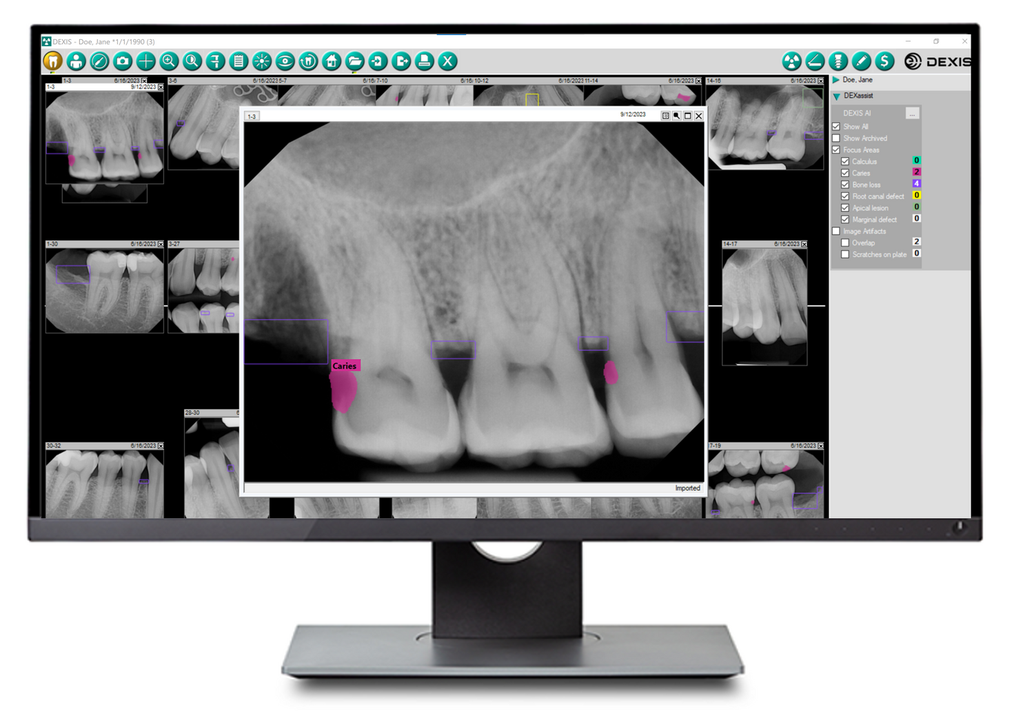
How to Interpret and Use This Output:
- Initial AI Analysis: The software flags two potential carious lesions and one area of calculus. This is your starting point. It has automatically screened the image and is drawing your attention to specific locations.
- Visual and Score Interpretation: You zoom in on the highlighted areas. You assess the shape and radiolucency of the flagged lesions. You note the confidence score (even if it's just a color code). Is it a high-confidence (very likely) or low-confidence (possible) finding?
- Clinical Correlation (Your Expertise): This is the most important step.
- You perform a visual exam of the patient's actual teeth. Is there visible cavitation or discoloration corresponding to the AI finding?
- You use your explorer to gently assess the texture. Is the surface hard and arrested, or soft and active?
- You consider the patient's history. Have they had caries here before? What is their overall risk profile?
- Synthesis and Final Diagnosis: You combine the AI's "suggestion" with your own clinical findings to make the final diagnosis. The AI might have correctly identified an incipient lesion that warrants placing on a "watch" list, or it may have confirmed a clear D2 lesion that requires restoration. The AI finding, especially with a high confidence score, serves as powerful evidence to support your diagnosis and to explain the need for treatment to the patient.
Conclusion
In this lesson, we peeled back the cover on AI outputs to understand what they truly represent. You've learned to interpret the language of AI software, from visual highlights to numerical scores, and place it in the proper clinical context.
Key Takeaways:
- AI systems use bounding boxes, segmentation masks, and highlighting to visually communicate their findings on a radiograph.
- A confidence score represents the model's certainty in its prediction based on pattern matching, not the absolute probability of disease.
- The clinician's role is to critically evaluate the AI's output, correlate it with clinical examination and patient history, and avoid over-reliance or "diagnostic anchoring."
- AI is an assistive tool that acts as a "second opinion" or an automated screener, augmenting—not replacing—your professional judgment.
Preview of the Next Lesson
You now know how to run an AI analysis and how to interpret the results. The next step is to formally bring it all together. In our next lesson, we will focus on how to integrate AI findings with clinical examination data to formulate a comprehensive diagnosis. We will work through case scenarios to practice this synthesis, moving from individual findings to a complete diagnostic picture.