Hello! Welcome back to your course on AI in dentistry.
In our last lesson, we focused on interpreting the specific outputs of AI software—the bounding boxes, segmentation masks, and confidence scores. We established that these are powerful assistive tools, but that you, the clinician, remain the ultimate diagnostician, responsible for critically evaluating the AI's suggestions and avoiding "diagnostic anchoring."
Today, we will take that a step further. The learning outcome for this lesson is to integrate AI findings with clinical examination data to formulate a comprehensive diagnosis. This is where the true power of AI is unlocked—not just as a detection tool, but as a component in a holistic diagnostic process. We will move from looking at individual AI flags to building a complete clinical picture.
1. The Synthesis Framework: From Data to Diagnosis
A robust diagnosis isn't based on a single piece of information. It's a synthesis of multiple data streams. When using AI, we can think of this as a three-part framework:
- AI Findings (The "What"): This is your starting point. The AI has automatically screened radiographs, scans, or photos and is drawing your attention to potential areas of interest. It might detect caries, measure bone loss, or segment a periapical lesion. This is the AI's contribution: objective, quantitative data derived from images.
- Clinical Examination (The "How"): This is where you provide the essential "ground truth." You take the AI's findings to the chairside and investigate. This includes your visual exam, tactile assessment with an explorer, periodontal probing, vitality testing, and palpation. The clinical exam confirms, refutes, or adds nuance to the AI's suggestions.
- Patient Context (The "Why"): This layer provides the narrative. It includes the patient's chief complaint, medical and dental history, habits (diet, hygiene), and risk factors. This context is crucial for moving from a simple finding (e.g., "radiolucency") to a diagnosis (e.g., "active carious lesion in a high-risk patient") and formulating an appropriate treatment plan.
A comprehensive diagnosis lies at the intersection of all three. The AI acts as a diligent assistant, ensuring nothing is missed, while you act as the expert, integrating all the evidence to make the final call.
The American Dental Association's white paper on AI touches on this integration, particularly how AI can cross-reference imaging data with electronic dental records (EDR) to support clinical decision-making.
[PDF] Overview of Artificial and Augmented Intelligence Uses in Dentistry
Let's revisit the ADA's 'Overview of Artificial and Augmented Intelligence Uses in Dentistry'. This time, focus on the section that explicitly discusses how AI helps connect different data sources.
Please read the subsection titled 'Electronic Dental Record (EDR) Use' (around page 22). Pay close attention to the paragraph under 'Diagnosis, Treatment Planning, and Risk Assessment.' It describes how AI can integrate and analyze various data types from the EDR to facilitate clinical decision-making.
Now, let's see how this framework applies in practice through a few case scenarios.
2. Case Scenario 1: Restorative Diagnosis
Imagine a patient is in for a routine check-up. You take bitewing radiographs, and the AI software provides the following analysis.
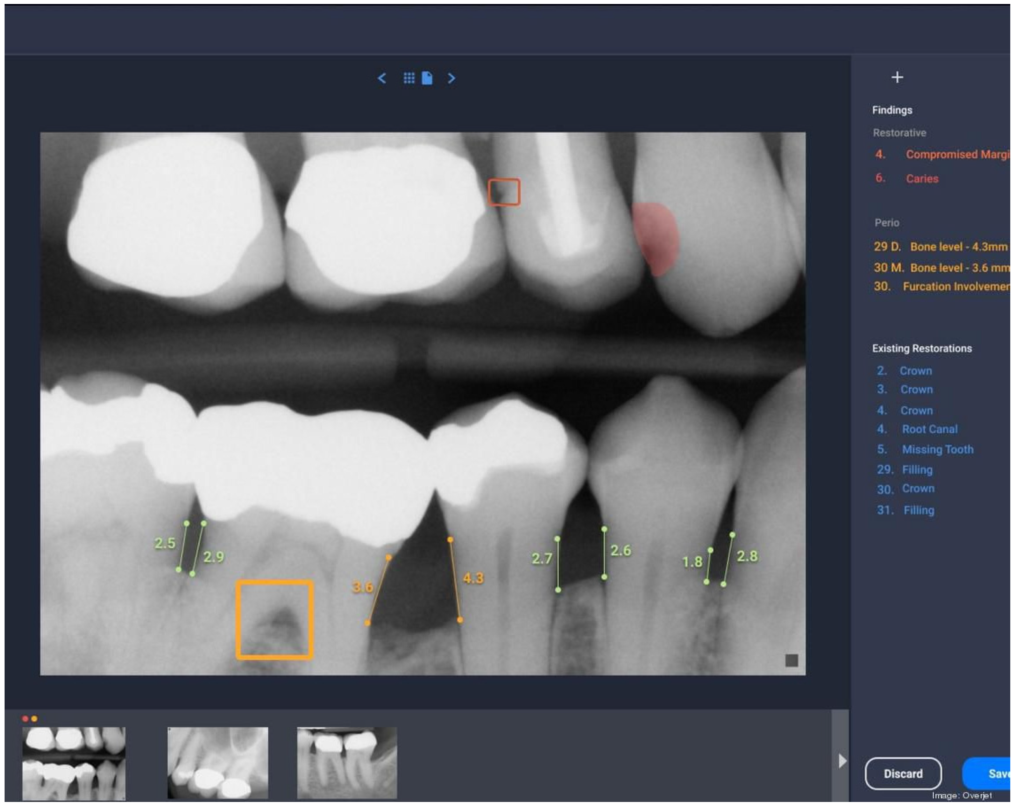
Let's apply our framework:
- AI Findings (The "What"): The AI highlights a radiolucency on the distal of the first premolar, labeling it as 'Caries' with high confidence. It also flags a potential open margin on the existing restoration on the second premolar.
- Clinical Examination (The "How"):
- You perform a visual exam. You can't see the interproximal lesion on the first premolar directly, but you notice a slight greyish discoloration on the marginal ridge.
- Using an explorer, you feel a slight "stick" in that interproximal area.
- Your examination of the second premolar confirms a detectable gap between the restoration and the tooth structure, which corresponds to the AI's 'Compromised Margin' finding.
- Patient Context (The "Why"): The patient mentions they don't floss regularly and have a sweet tooth. Their record shows they've had several fillings in the past.
Synthesis and Comprehensive Diagnosis:
The AI's findings are strongly corroborated by your clinical exam and the patient's risk profile.
- Diagnosis for the first premolar: Active interproximal carious lesion.
- Diagnosis for the second premolar: Defective restoration with a compromised margin, indicating a risk for recurrent decay.
Here, the AI didn't tell you something you couldn't have found yourself, but it streamlined the process. It drew your eye immediately to the problem areas, and its visual output is now an excellent tool for educating the patient about why treatment is necessary.
3. Case Scenario 2: Periodontal Diagnosis
Next, consider a patient presenting with concerns about "bleeding gums." The AI analyzes their full mouth series of radiographs and periodontal charting data.

- AI Findings (The "What"): The AI has measured radiographic bone loss (RBL) across the dentition, flagging several teeth with RBL exceeding 30%. It also integrates with the periodontal chart to highlight a 5mm pocket on tooth #18.
- Clinical Examination (The "How"):
- You perform full-mouth periodontal probing. Your measurements confirm 5mm and 6mm pockets on the molars, including #18, with generalized bleeding on probing (BOP).
- You assess tooth mobility and find Class I mobility on tooth #18.
- Patient Context (The "Why"): The patient is a 50-year-old with type 2 diabetes and a history of smoking. They admit to being overdue for a cleaning.
Synthesis and Comprehensive Diagnosis:
The AI's quantitative analysis of bone loss provides an objective measure that aligns perfectly with your clinical findings of deep pockets and BOP. The patient's systemic risk factors (diabetes, smoking) complete the picture.
- Diagnosis: Generalized Periodontitis, Stage III, Grade C.
In this case, the AI excels at quantification and tracking. It provides a precise, objective baseline of bone levels that can be compared at future appointments to monitor disease progression or stability after treatment.
4. Case Scenario 3: Comprehensive Treatment Planning
AI's integrative power shines in complex cases that involve multiple data types, such as aesthetic and functional rehabilitations.
Smilecloud Tutorial 3DNA | AI Smile Design and Cloud Dentistry
This video from Digital Dentistry Masterclass demonstrates SmileCloud, a platform that uses AI to integrate various patient data for comprehensive smile design and treatment planning. It's a fantastic example of synthesis in action.
Please watch the following segments: Case Setup (2:29 - 4:09): Notice how different data types are uploaded—intraoral scans, photos, CBCT, and even jaw motion data. AI Smile Design (5:16 - 7:22): See how the AI uses a 2D photo to generate an initial smile design proposal. Creating the 3D Blueprint (7:22 - 9:36): This is the key part. Watch how the 2D design is merged with 3D data (scans, CBCT) to create a comprehensive 'digital patient.' This allows for analysis of aesthetics, function, and underlying structures all at once.
Let's break this down using our framework:
- AI Findings (The "What"):
- The AI uses facial biometrics from a photo to propose an ideal smile design (tooth proportions, smile line).
- It then automatically segments the teeth and bone from the CBCT scan.
- It merges the intraoral scans, CBCT, and photo to create a photorealistic 3D avatar.
- It uses motion data to display dynamic occlusal contacts on a heatmap.
- Clinical Examination (The "How"):
- Your intraoral exam confirms the presence of worn incisal edges, malpositioned teeth, and occlusal discrepancies that the AI's data has visualized.
- A functional analysis confirms the patient has a constricted chewing pattern, explaining some of the wear.
- Patient Context (The "Why"): The patient's chief complaint is purely aesthetic: "I don't like my smile."
Synthesis and Comprehensive Treatment Plan:
The AI-driven platform doesn't just give a diagnosis; it facilitates a comprehensive treatment plan.
- Diagnosis: Malocclusion and tooth wear with aesthetic concerns.
- Integration for Planning: You, the clinician, use the integrated 3D avatar to make critical decisions. You can see that simply placing veneers (an aesthetic-only fix) would fail due to the underlying functional issues. The platform allows you to simulate different options, such as orthodontics followed by restorative work. You can design the final tooth shapes and verify that they will function correctly during chewing movements before you ever touch the patient's mouth. The diagnosis and treatment plan become one fluid, digitally-planned process.
5. The Ultimate Check: Your Professional Judgment
What happens when your clinical exam contradicts the AI? For example, the AI flags a dark spot on a radiograph as "potential caries," but your exam reveals it's a non-carious stain or a misleading shadow from tooth overlap.
This is where your role is most critical. The clinician's judgment always supersedes the AI's output. AI is an assistive tool, not an oracle. Professional regulatory bodies are very clear about this responsibility.
Information on Artificial Intelligence
The Royal College of Dental Surgeons of Ontario (RCDSO) provides clear guidance on this exact issue. Let's review their stance on dentist accountability.
Please read the answers to the following three questions: 'What happens if dentists disagree with an AI-generated output?' 'Are dentists responsible if using AI leads to an error?' 'What is important for dentists to document when using AI?' Focus on the core message about exercising professional judgment, your ultimate responsibility for care, and the importance of documenting your rationale.
The key takeaway is that you are expected to critically review and, when necessary, override the AI's findings. Documenting your decision—whether you agree or disagree with the AI and why—is a crucial part of integrating this technology into the patient record.
Conclusion
In this lesson, you've learned a systematic framework for moving beyond simply looking at AI outputs to truly integrating them into your diagnostic workflow.
Key Takeaways:
- A comprehensive diagnosis synthesizes AI findings, clinical examination data, and patient context.
- AI excels at automated screening, quantification, and integrating multiple digital data sources, serving as a powerful starting point.
- Your clinical examination provides the essential ground truth to confirm or refute the AI's suggestions.
- Your professional judgment is paramount. You are responsible for the final diagnosis and must be prepared to override AI findings when they contradict your clinical assessment.
- Properly integrated, AI doesn't just help find pathology; it helps build more robust, evidence-based, and comprehensive treatment plans.
Preview of the Next Lesson
Now that you know how to formulate a comprehensive, AI-assisted diagnosis, the next logical step is sharing that information. In our next lesson, we will focus on how to formulate clear and ethical communication strategies for discussing AI-assisted findings with patients. You'll learn how to leverage the visual power of AI to enhance patient education, build trust, and improve treatment acceptance.