Welcome to our next lesson in neuroanatomy. In the previous session, we explored the cerebellum's function as the master coordinator of movement, learning to distinguish between midline and hemispheric lesions based on their classic signs like truncal versus limb ataxia.
Today, we transition from the subcortical motor systems to the peripheral connections of the brain itself. We will begin our systematic study of the twelve cranial nerves, which are absolutely fundamental for your MPT entrance exams. This lesson focuses on the first six nerves, Cranial Nerves I through VI. We will identify their specific functions, the clinical tests used to assess them, and the hallmark signs that appear when they are damaged. Mastering this content is crucial, as questions on cranial nerve palsies are a staple of neuro-physiotherapy.
An Overview of the Cranial Nerves
The twelve pairs of cranial nerves emerge directly from the brain and brainstem, providing vital sensory and motor connections to the head, neck, and various visceral organs. Before we dive into the first six individually, let's visualize where they originate.
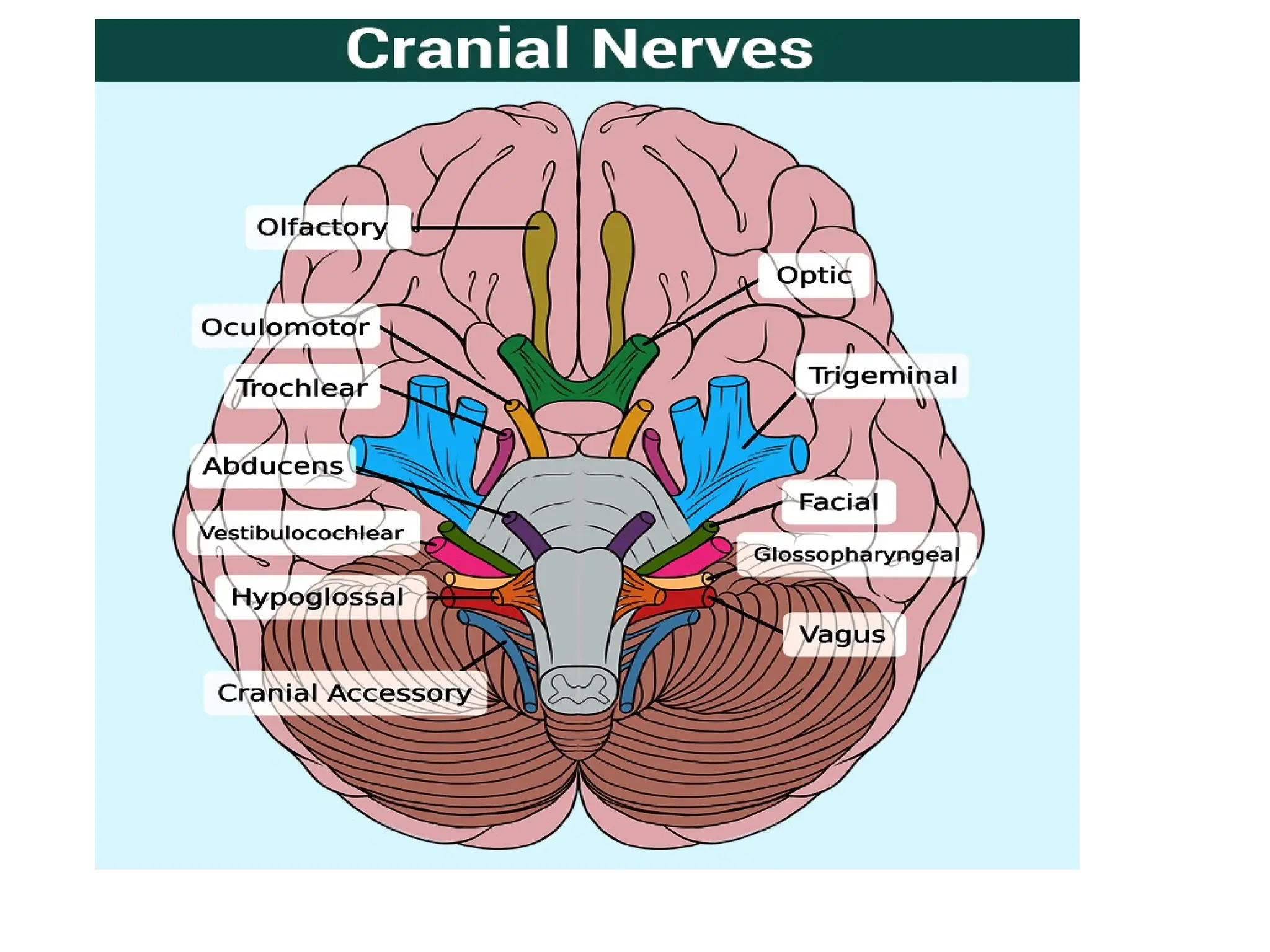
This superior view of the brain and brainstem shows the points of exit for the twelve cranial nerves. Notice how most of them emerge from the brainstem (midbrain, pons, and medulla).
For exam purposes, it's essential to memorize the names of the nerves, their order, and whether their primary function is sensory (S), motor (M), or both (B). Two popular mnemonics will help you with this:
- For the names (CN I to XII): "On Old Olympus' Towering Top, A Finn And German Viewed Some Hops."
- (Olfactory, Optic, Oculomotor, Trochlear, Trigeminal, Abducens, Facial, Auditory/Vestibulocochlear, Glossopharyngeal, Vagus, Spinal Accessory, Hypoglossal)
- For the function (S, M, or B): "Some Say Marry Money, But My Brother Says Big Brains Matter More."
Let's begin our detailed exploration, nerve by nerve.
CN I: Olfactory Nerve
- Function: Special Sensory (Smell)
- Lesion Sign: Anosmia (loss of smell)
The olfactory nerve is purely sensory and conveys the sense of smell. Its fibers originate from receptors in the nasal mucosa, pass through the cribriform plate of the ethmoid bone, and synapse in the olfactory bulb.
Clinical Testing: The test is straightforward. With the patient's eyes closed, you occlude one nostril and ask them to identify a common, non-irritating scent like coffee or cinnamon. You then repeat the test on the other side.
Cranial Nerves Exam | Clinical Skills
The Ninja Nerd video provides a clear clinical demonstration of how to test the olfactory nerve.
Watch the segment from the start of the CN I test. Notice the initial step of checking for nasal patency before presenting the smells. The video also lists common causes of anosmia, such as head trauma (fracture of the cribriform plate), viral infections (like COVID-19), and tumors.
CN II: Optic Nerve
- Function: Special Sensory (Vision)
- Lesion Sign: Varies from visual field defects to complete blindness.
The optic nerve is also purely sensory, transmitting visual information from the retina to the brain. Assessing this nerve is more complex and involves several components.
Cranial Nerve Examination - OSCE Guide | Geeky Medics
The Geeky Medics guide provides a thorough, step-by-step written explanation of how to assess the optic nerve, which is an excellent reference to complement the video demonstration.
Please read the section on the Optic Nerve. Pay close attention to the descriptions and procedures for: Visual acuity (Snellen chart) Pupillary reflexes Visual fields (confrontation testing) and the different types of field defects.
Let's break down the key tests for the optic nerve:
- Visual Acuity: This tests the sharpness of central vision using a Snellen chart. It's recorded as a fraction (e.g., 6/6 or 20/20).
- Visual Fields: This assesses peripheral vision. The confrontation test, where you compare the patient's visual field to your own, is standard. Lesions along the visual pathway cause characteristic field defects (e.g., a pituitary tumor compressing the optic chiasm causes bitemporal hemianopia).
- Pupillary Light Reflex: This tests both CN II (the afferent limb, sensing the light) and CN III (the efferent limb, constricting the pupil). When you shine a light in one eye, both that eye (direct reflex) and the opposite eye (consensual reflex) should constrict.
- Swinging Light Test: This is a crucial test to detect a relative afferent pupillary defect (RAPD), also known as a Marcus Gunn pupil. An RAPD indicates optic nerve disease (e.g., optic neuritis). When light is swung from the normal eye to the affected eye, the affected eye's pupil paradoxically dilates instead of constricting because it senses less light.
- Fundoscopy: Using an ophthalmoscope to visualize the back of the eye, particularly the optic disc. A swollen disc (papilledema) is a sign of raised intracranial pressure.
Cranial Nerves Exam | Clinical Skills
This video demonstrates the full sequence of testing the optic nerve, bringing the written guide to life.
Watch the comprehensive segment on the optic nerve exam. This covers visual acuity, the finger counting method for visual fields, pupillary reflexes including the swinging light test, and fundoscopy.
The Eye Movers: CN III, IV, and VI
These three nerves are best learned together as they coordinate all movements of the eye. A simple mnemonic helps you remember which nerve supplies which muscle: LR₆(SO₄)₃.
- LR₆: The Lateral Rectus is supplied by CN VI (Abducens).
- SO₄: The Superior Oblique is supplied by CN IV (Trochlear).
- ₃: All the rest of the extraocular muscles are supplied by CN III (Oculomotor).
Clinical Testing: The primary test is to assess extraocular movements. You ask the patient to keep their head still and follow your finger as you trace a large "H" pattern. This takes their eyes through all cardinal directions of gaze. During this, you watch for any limitation of movement and ask the patient if they experience any double vision (diplopia).
CN III: Oculomotor Nerve
- Function: Motor to the superior, inferior, and medial recti, the inferior oblique, and the levator palpebrae superioris (lifts the eyelid). It also carries parasympathetic fibers that constrict the pupil and control accommodation.
- Lesion Sign: A complete CN III palsy results in a classic triad:
- The eye is positioned "down and out" due to the unopposed action of the lateral rectus (CN VI) and superior oblique (CN IV).
- Ptosis (drooping eyelid) because the levator palpebrae is paralyzed.
- A fixed, dilated pupil that doesn't react to light or accommodation.
CN IV: Trochlear Nerve
- Function: Motor to the superior oblique muscle, which moves the eye downward and inward (intorsion).
- Lesion Sign: The patient has difficulty looking down and in. This causes vertical diplopia, which is often most pronounced when walking down stairs or reading. Patients typically compensate by tilting their head away from the side of the lesion.
CN VI: Abducens Nerve
- Function: Motor to the lateral rectus muscle, which abducts (moves outward) the eye.
- Lesion Sign: The patient cannot abduct the affected eye. At rest, the eye deviates medially, creating a convergent squint (esotropia). This causes horizontal diplopia that worsens when they try to look towards the side of the lesion.
Cranial Nerves Exam | Clinical Skills
This part of the video demonstrates the "H" pattern test for extraocular movements and discusses the functions of these three nerves.
Watch the section covering CN III, IV, and VI. The video explains the LR6(SO4)3 mnemonic and shows how to check for ptosis.
CN V: Trigeminal Nerve
- Function: Mixed (Both Sensory and Motor)
- Lesion Sign: Sensory loss on the face; weakness/wasting of mastication muscles.
The trigeminal nerve is the principal sensory nerve for the face and also controls the muscles of chewing.
- Sensory Component: Supplies sensation to the face via three divisions:
- V₁ (Ophthalmic): Forehead and scalp, upper eyelid, cornea.
- V₂ (Maxillary): Cheek, lower eyelid, upper lip, upper teeth.
- V₃ (Mandibular): Jaw, chin, lower lip, lower teeth, general sensation (not taste) from the anterior 2/3 of the tongue.
- Motor Component: Innervates the muscles of mastication (temporalis, masseter, pterygoids).
Clinical Testing:
- Sensory: With the patient's eyes closed, test light touch (cotton wool) and pinprick sensation over the three divisions, comparing both sides.
- Motor: Ask the patient to clench their jaw and palpate the bulk and strength of the temporalis and masseter muscles. Then, ask them to open their mouth against resistance to test the pterygoids. In a unilateral lesion, the jaw will deviate towards the weak side upon opening.
- Reflexes:
- Corneal Reflex: Touching the cornea with a wisp of cotton wool should cause both eyes to blink. The afferent limb is CN V (V₁), and the efferent limb is CN VII (Facial nerve).
- Jaw Jerk Reflex: Tapping the chin with the mouth slightly open causes a slight closure. This reflex (involving both afferent and efferent CN V fibers) is often absent in healthy individuals but becomes brisk in upper motor neuron lesions.
Cranial Nerve Examination - OSCE Guide | Geeky Medics
This section of the Geeky Medics guide details the sensory and motor testing for the trigeminal nerve, including the reflexes.
Read the section on the Trigeminal Nerve. Focus on the specific areas to test for each of the three sensory divisions and the procedure for testing the muscles of mastication.
High-Yield Summary for Exam Revision
This cheat sheet provides a fantastic, quick-reference summary of the functions and assessments for all cranial nerves. For this lesson, focus on the first six.

This Cranial Nerves Assessment Cheat Sheet summarizes the name, function, and assessment method for each nerve. It is an excellent tool for rapid revision before your exam.
| Nerve | Function(s) | Key Test(s) | Classic Lesion Sign(s) |
|---|---|---|---|
| I. Olfactory | Smell (Sensory) | Identify common odors | Anosmia (loss of smell) |
| II. Optic | Vision (Sensory) | Acuity, Visual Fields, Pupillary Reflexes | Field defects, decreased acuity, RAPD |
| III. Oculomotor | Eye movement, Pupil constriction (Motor) | "H" pattern, Pupillary light reflex | Eye "down and out", Ptosis, Dilated pupil |
| IV. Trochlear | Eye movement (down & in) (Motor) | "H" pattern | Vertical diplopia, Head tilt away from lesion |
| V. Trigeminal | Facial sensation, Mastication (Both) | Light touch, Pinprick, Jaw clench/opening | Facial numbness, Jaw deviates towards lesion |
| VI. Abducens | Eye abduction (Motor) | "H" pattern | Inability to abduct eye, Horizontal diplopia |
Conclusion
In this lesson, we have systematically covered the first six cranial nerves, which are critical for sensory input from the face and for precise control of eye movements. You have learned their individual functions, how to perform key clinical tests, and how to recognize the classic signs of their dysfunction.
Key Takeaways:
- Cranial nerves provide the direct neural link between the brain and the head/neck.
- CN I (Olfactory) is for smell; its loss is anosmia.
- CN II (Optic) is for vision; its assessment involves acuity, fields, and pupillary reflexes. An RAPD points to optic nerve pathology.
- The eye movement nerves CN III, IV, and VI work together. Remember their classic palsies: CN III ("down and out," ptosis, blown pupil), CN IV (vertical diplopia, head tilt), and CN VI (medial squint, cannot abduct).
- CN V (Trigeminal) has three sensory branches for the face and a motor branch for chewing.
In our next lesson, we will continue our journey through the cranial nerves by examining nerves VII through XII. These include the facial nerve, the vestibulocochlear nerve (for hearing and balance), and the nerves that control the intricate movements of your palate, pharynx, and tongue.