Welcome back to our lessons on neuroanatomy. In our previous session, we established a solid foundation by examining the first six cranial nerves, covering their roles in smell, vision, eye movement, and facial sensation. You learned to identify the classic signs of their dysfunction, such as the "down and out" eye in an oculomotor nerve palsy.
Today, we will complete our survey of the twelve cranial nerves by focusing on nerves VII through XII. This lesson is critical for your MPT exam preparation, as these nerves govern functions you frequently assess and treat as a physiotherapist: facial expression, hearing and balance, swallowing, speech, and movements of the neck and tongue. We will identify their functions, the standard clinical tests for each, and the key signs of lesions, paying special attention to high-yield facts for your upcoming exams.
CN VII: Facial Nerve
- Function: Mixed (Both Sensory and Motor)
- Lesion Signs: Unilateral facial weakness/paralysis, altered taste, hyperacusis.
The facial nerve is arguably one of the most clinically significant cranial nerves in physiotherapy. Its primary role is motor, supplying all the muscles of facial expression. It also carries taste sensation from the anterior two-thirds of the tongue and provides parasympathetic innervation to the lacrimal and submandibular/sublingual salivary glands.
Clinical Testing
Assessment involves observing the face for symmetry and then testing the strength of specific facial muscles.
Cranial Nerve Examination - OSCE Guide | Geeky Medics
The Geeky Medics guide provides a clear, step-by-step protocol for assessing the facial nerve.
Please read the section on the Facial nerve. Focus on the sequence of motor assessment commands: Inspect for asymmetry at rest (e.g., drooping mouth corner, flattened nasolabial fold). Raise eyebrows (frontalis). Close eyes tightly and resist opening (orbicularis oculi). Show teeth or smile (multiple muscles). Puff out cheeks (buccinator).
Differentiating Upper vs. Lower Motor Neuron Lesions
This distinction is a classic topic for exams and is essential for clinical reasoning. A lesion's location determines the pattern of facial weakness.
- Lower Motor Neuron (LMN) Lesion: This involves the facial nerve nucleus or the nerve itself after it has left the brainstem. It causes paralysis of the entire ipsilateral (same side) half of the face. The patient cannot wrinkle their forehead or close their eye on the affected side. The most common example is Bell's Palsy.
- Upper Motor Neuron (UMN) Lesion: This involves the corticobulbar tracts that run from the motor cortex to the brainstem. Because the upper part of the facial nucleus (which controls the forehead) receives bilateral innervation from the cortex, the forehead is spared. This results in paralysis of the contralateral (opposite side) lower face only. The patient can still wrinkle their forehead. A common cause is a stroke.
This diagram clearly illustrates the difference in presentation.
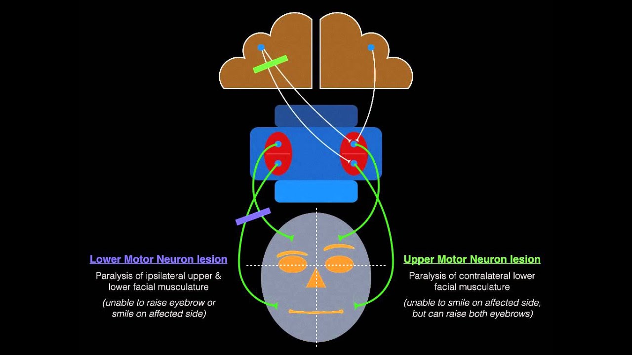
This diagram contrasts the effects of a Lower Motor Neuron (LMN) lesion and an Upper Motor Neuron (UMN) lesion on facial musculature. Note the forehead sparing in the UMN lesion.
Cranial Nerves Exam | Clinical Skills
The Ninja Nerd video provides an excellent clinical demonstration of the tests for the facial nerve and reinforces the UMN vs. LMN concept.
Watch the segment covering the facial nerve exam. Pay close attention to the visual checks for asymmetry and the series of commands given to the patient.
CN VIII: Vestibulocochlear Nerve
- Function: Special Sensory
- Lesion Signs: Hearing loss, tinnitus, vertigo, nystagmus.
This nerve has two distinct components: the cochlear nerve for hearing and the vestibular nerve for balance and spatial orientation.
Clinical Testing
-
Cochlear (Hearing):
- Gross Hearing: A simple screen is done by rubbing your fingers together by each ear or using a whispered voice.
- Rinne's Test: Compares air conduction (AC) to bone conduction (BC). A tuning fork is placed on the mastoid process (BC) and then moved next to the ear canal (AC). Normally, AC > BC.
- Weber's Test: A tuning fork is placed on the midline of the forehead. Normally, the sound is heard equally in both ears. In conductive hearing loss, it lateralizes to the bad ear; in sensorineural loss, it lateralizes to the good ear.
-
Vestibular (Balance):
- Tests like the Head Thrust Test or observing for nystagmus are used to assess the vestibulo-ocular reflex, but a detailed vestibular assessment is typically reserved for patients with specific complaints of dizziness or imbalance.
Cranial Nerve Examination - OSCE Guide | Geeky Medics
The Geeky Medics guide provides one of the clearest explanations of how to perform and interpret Rinne's and Weber's tests.
Read the section on the Vestibulocochlear nerve, focusing on the detailed procedures for the gross hearing assessment, Rinne's test, and Weber's test. The distinction between conductive and sensorineural hearing loss is a high-yield topic.
CN IX (Glossopharyngeal) & CN X (Vagus)
These two nerves are often tested together due to their overlapping functions in the pharynx and larynx.
- CN IX (Glossopharyngeal): Mixed function. Provides taste and general sensation to the posterior 1/3 of the tongue, innervates the stylopharyngeus muscle (aids swallowing), and is the afferent (sensory) limb of the gag reflex.
- CN X (Vagus): Mixed function. Provides motor supply to the muscles of the palate, pharynx, and larynx. It is the efferent (motor) limb of the gag reflex. It also has extensive autonomic functions throughout the thorax and abdomen.
Clinical Testing & Lesion Signs
- Voice Quality: Ask the patient to speak. A hoarse or "breathy" voice can indicate a vagus nerve lesion affecting the vocal cords.
- Palate Elevation: Ask the patient to open their mouth and say "Ahhh."
- Observation: Look at the soft palate and uvula.
- Normal: The palate should elevate symmetrically, and the uvula should remain in the midline.
- Lesion: In a unilateral vagus nerve (CN X) lesion, the palate on the affected side will not rise, and the uvula will deviate AWAY from the side of the lesion.
- Swallowing: Ask the patient to take a small sip of water and observe for any difficulty (dysphagia) or coughing.
- Gag Reflex: Touching the back of the pharynx should cause gagging (afferent IX, efferent X). This is unpleasant and often deferred, but you must know the pathways.
CN XI: Accessory Nerve
- Function: Motor
- Lesion Sign: Weakness of shoulder shrug and head rotation.
This nerve is unique as it originates from the cervical spinal cord, ascends into the skull, and then exits to supply two key muscles: the sternocleidomastoid (SCM) and the upper trapezius.
Clinical Testing
- Trapezius: Ask the patient to shrug their shoulders against your resistance. Look for weakness or asymmetry. A lesion can cause shoulder drooping.
- Sternocleidomastoid: Ask the patient to turn their head to one side against your resistance. The SCM contracts to turn the head to the opposite side.
CN XII: Hypoglossal Nerve
- Function: Motor
- Lesion Sign: Tongue deviation, atrophy, fasciculations.
This nerve provides motor innervation to all the intrinsic and extrinsic muscles of the tongue (except palatoglossus, by CN X).
Clinical Testing
- Inspection: Ask the patient to open their mouth and inspect the tongue at rest for any signs of wasting (atrophy) or fine, rippling movements (fasciculations), which are signs of an LMN lesion.
- Protrusion: Ask the patient to stick their tongue straight out.
- Observation: Note any deviation.
- Lesion: In a unilateral CN XII lesion, the tongue will deviate TOWARDS the side of the lesion. The mnemonic is "you lick your wounds." This happens because the unopposed action of the genioglossus muscle on the healthy side pushes the tongue towards the weak side.
Cranial nerves IX, X, XI & XII
This short video from The Noted Anatomist provides a very clear, anatomically-focused overview of the last four cranial nerves. It's a great way to consolidate your understanding of their functions before reviewing the clinical tests.
Watch the entire video (CN IX, X, XI, & XII) to quickly review the origin, course, and primary functions of each of these four nerves.
A guide to cranial nerve testing for musculoskeletal clinicians - PMC
This article provides concise, clinically-oriented descriptions of testing and lesion mechanisms, which is perfect for your level of study.
First, review the sections covering the tests for these four nerves (CN VII, IX, X, XII, and XI). Then, jump to the appendix and read the "Mechanisms" section for each of the nerves from CN VII to CN XII. This will give you a deeper understanding of the pathology behind the signs you observe.
High-Yield Summary for Exam Revision
As you prepare for your exam, being able to quickly recall the key features of each nerve is essential. This infographic provides a fantastic overview of all twelve nerves. Use it as a self-testing tool.

This infographic summarizes the functions, tests, and mnemonics for all twelve cranial nerves. It is an excellent tool for quick revision.
| Nerve | Function(s) | Key Test(s) | Classic Lesion Sign(s) |
|---|---|---|---|
| VII. Facial | Facial expression, Taste (ant. 2/3) | Raise eyebrows, smile, puff cheeks | LMN: Full ipsilateral paralysis. UMN: Contralateral lower face paralysis (forehead spared). |
| VIII. Vestibulocochlear | Hearing, Balance | Finger rub, Rinne/Weber tests | Hearing loss, tinnitus, vertigo, nystagmus. |
| IX. Glossopharyngeal | Taste (post. 1/3), Swallowing, Gag (sensory) | Gag reflex, swallowing | Loss of taste (post.), absent gag reflex. |
| X. Vagus | Swallowing, Phonation, Gag (motor) | Say "Ahhh", swallow, assess voice | Hoarseness, dysphagia, uvula deviates AWAY from lesion. |
| XI. Accessory | Shoulder shrug, Head rotation | Resisted shoulder shrug, head turn | Shoulder droop, weakness in shrug & head rotation. |
| XII. Hypoglossal | Tongue movement | Protrude tongue | Tongue deviates TOWARDS lesion, atrophy, fasciculations. |
Conclusion
In this lesson, we have completed our foundational tour of the cranial nerves. You have learned the functions, clinical tests, and lesion signs for nerves VII through XII, which are integral to facial movement, communication, swallowing, and head control.
Key Takeaways:
- Differentiating UMN and LMN facial nerve lesions is crucial: UMN spares the forehead, while LMN affects the entire hemiface.
- Rinne's and Weber's tests are used to differentiate conductive from sensorineural hearing loss (CN VIII).
- The uvula deviates away from a CN X lesion.
- The tongue deviates towards a CN XII lesion.
- CN XI weakness presents as a drooping shoulder and difficulty with head rotation.
We have now examined the major anatomical "wiring" of the central and peripheral nervous systems. In our next module, we will shift our focus from neuroanatomy to neurophysiology. We will begin by exploring the fundamental unit of neural communication: the action potential. Understanding how neurons generate and propagate electrical signals is the key to comprehending everything from sensory perception to motor commands.