Welcome to our next lesson in neuroanatomy. In our previous session, we examined the basal ganglia's critical role in motor control, focusing on how the direct and indirect pathways act as a "Go" and "No-Go" system to select appropriate movements. We saw how imbalances in this system lead to the hypokinetic signs of Parkinson's disease and the hyperkinetic features of Huntington's disease.
Today, we shift our focus from selecting a movement to perfecting it. We will explore another major motor control center: the cerebellum. This lesson will cover the anatomy and primary functions of the cerebellum and, most importantly for your clinical practice and exam preparation, teach you how to differentiate between the various types of cerebellar lesions based on their distinct clinical presentations. This topic is fundamental to neurological physiotherapy and is frequently tested in MPT entrance exams.
The Cerebellum: The Brain's Quality Control Manager
While the basal ganglia help decide which motor program to run, the cerebellum ensures the program runs smoothly, accurately, and efficiently. You can think of it as a quality control manager for movement. It doesn't initiate movement itself, but it constantly compares the intended movement (the "plan" from the cerebral cortex) with the actual movement (the "feedback" from proprioceptors in your muscles and joints) and issues real-time corrections.
This function is vital for a huge range of activities, from the precise finger movements of typing to maintaining your balance while walking on an uneven surface.
To begin, let's get an overview of the cerebellum's structure and its principal functions.
This Physiopedia article provides a concise introduction to the cerebellum's location, its primary functions in motor control, and its basic structure.
Please read the first three main sections. Start with the overview and functions to get a sense of its role. Then, continue to the section on Cerebellar Structure, focusing on the general layout of the hemispheres, vermis, and the grey/white matter.
Functional Anatomy: Mapping Function to Structure
To understand cerebellar disorders, you must first understand how the cerebellum is organized functionally. While it has anatomical lobes (anterior, posterior, flocculonodular), it's more clinically useful to divide it into three functional zones. Each zone has distinct connections and is responsible for a different aspect of motor control.

This diagram illustrates the main functional divisions of the cerebellum. From medial to lateral, these are the Vermis, Intermediate Zone, and Lateral Hemisphere, along with the Flocculonodular Lobe at the bottom. Each is linked to a primary motor function.
Let's break down these three functional divisions:
-
Vestibulocerebellum (Archicerebellum):
- Anatomy: Consists of the flocculonodular lobe.
- Function: This is the oldest part of the cerebellum. Its primary job is to process information from the vestibular system to control balance and eye movements (like the vestibulo-ocular reflex, which keeps your eyes fixed on a target when your head moves).
- Lesion Signs: Damage here leads to dysequilibrium, truncal ataxia (instability of the core), and nystagmus (involuntary eye movements).
-
Spinocerebellum (Paleocerebellum):
- Anatomy: Comprises the central vermis and the adjacent intermediate (paravermal) zones of the hemispheres.
- Function: It receives extensive proprioceptive input from the spinal cord (hence "spino-") and is responsible for coordinating movements of the trunk and limbs. The vermis specifically controls axial and truncal muscles, while the intermediate zone governs distal limb muscles. It also regulates muscle tone.
- Lesion Signs: A lesion in the vermis causes gait ataxia (a wide-based, unsteady, "drunken" gait). A lesion in the intermediate zone causes ataxia in the limbs (e.g., arms and legs).
-
Cerebrocerebellum (Neocerebellum):
- Anatomy: Made up of the large lateral hemispheres.
- Function: This is the newest part evolutionarily. It has extensive connections with the cerebral cortex (hence "cerebro-") and is crucial for the planning, timing, and coordination of skilled, voluntary movements. It's essential for learning new motor skills.
- Lesion Signs: Damage here results in a cluster of deficits affecting skilled limb movements, including dysmetria, dysdiadochokinesia, and intention tremor.
The following video from Armando Hasudungan provides a fantastic visual summary of these anatomical and functional divisions.
Cerebellum Clinical Anatomy - cerebellar syndrome
This animated video clearly illustrates the anatomical and functional organization of the cerebellum, which is key to understanding the different clinical syndromes.
Watch the first part of the video covering the cerebellum's anatomy. Pay close attention to how it maps the three functional areas (Cerebrocerebellum, Spinocerebellum, Vestibulocerebellum) to their corresponding anatomical parts and functions.
Clinical Signs of Cerebellar Dysfunction: The "DANISH" Mnemonic
When the cerebellum is damaged, the result is a loss of coordination known as ataxia. Unlike weakness (paresis) or a movement selection problem (as in basal ganglia disorders), ataxia is a problem of smoothness, timing, and accuracy.
A critical point for your exams and clinical practice is that cerebellar lesions cause deficits on the ipsilateral (same) side of the body. This is because the motor pathways that the cerebellum influences either don't cross or cross twice (effectively cancelling out the crossing). This is in stark contrast to the cerebral motor cortex, where a lesion causes contralateral deficits.
The common signs of cerebellar dysfunction can be remembered with the helpful mnemonic DANISH:
- D – Dysdiadochokinesia: Inability to perform rapid, alternating movements (e.g., rapidly pronating and supinating the forearm). The movement becomes slow, clumsy, and irregular.
- A – Ataxia: Uncoordinated movement. This can manifest as gait ataxia (a staggering, wide-based walk) or limb ataxia (clumsiness in the arms and legs).
- N – Nystagmus: Involuntary, rapid, oscillating eye movements.
- I – Intention Tremor: A tremor that appears during a voluntary, purposeful movement and worsens as the target is approached. This is very different from the resting tremor of Parkinson's disease.
- S – Slurred or Scanning Speech (Dysarthria): Speech is slow, disjointed, and syllables may be separated by pauses, as the coordination of speech muscles is impaired.
- H – Hypotonia: Decreased muscle tone. The muscles feel floppy and offer less resistance to passive movement.
This next section of the same video gives an excellent demonstration and explanation of these signs.
Cerebellum Clinical Anatomy - cerebellar syndrome
This part of the video explains the classic clinical signs of cerebellar damage using the "DANISH" acronym.
Watch the segment on clinical relevance. Focus on the explanation for each letter of the DANISH mnemonic and the demonstration of the test for dysdiadochokinesia. Note the crucial point made about ipsilateral deficits.
Differentiating Cerebellar Lesion Types: An Exam-Oriented Approach
For your MPT entrance exam, you'll need to be able to identify the location of a cerebellar lesion based on a patient's symptoms. The key is to map the signs back to the functional divisions we discussed.
1. Midline Lesions (Vermis & Flocculonodular Lobe)
These lesions primarily affect the spinocerebellum and vestibulocerebellum. The patient's core stability and gait are most affected.
- Key Signs: Truncal ataxia (difficulty sitting or standing without support), a wide-based "drunken" gait, titubation (a tremor of the head or trunk), and nystagmus. Limb coordination (e.g., finger-to-nose testing) might be surprisingly intact.
- Common Causes: Medulloblastoma (a common childhood tumor of the vermis), and atrophy due to chronic alcoholism.
2. Hemispheric Lesions (Lateral Hemispheres)
These lesions affect the cerebrocerebellum and the intermediate zone of the spinocerebellum. The deficits will be seen in the limbs on the same side as the lesion.
- Key Signs: Ipsilateral limb (appendicular) ataxia. This includes:
- Dysmetria: Inability to judge distance, leading to over- or undershooting a target (e.g., in a finger-to-nose test).
- Dysdiadochokinesia.
- Intention tremor.
- Dysarthria.
- Common Causes: Stroke (e.g., in the PICA or SCA arteries), tumor, multiple sclerosis plaque, or an abscess.
To solidify this, please read the following text, which clearly distinguishes between these syndromes.
Neuroanatomy, Cerebellum - StatPearls - NCBI Bookshelf - NIH
This text from StatPearls provides a clinically-focused summary of cerebellar signs and differentiates between vermis and cerebellar hemisphere syndromes.
In the "Clinical Significance" section, first read the paragraph that describes the classic signs, from hypotonia and ataxia to dysdiadochokinesia. Then, focus on the next paragraph that distinguishes between the two main syndromes, starting here.
For a comprehensive review of specific conditions affecting the cerebellum that are high-yield for exams, this video lecture is an excellent resource.
Cerebellum Anatomy and Clinical Syndromes
This lecture by Dr. Brad Cole provides a deeper dive into the functional lobes and a range of clinical conditions that cause cerebellar ataxia.
First, watch the segment on the functional lobes for a reinforcement of how lesions in the anterior, posterior, and flocculonodular lobes produce different types of ataxia. Then, watch the section on clinical conditions. Pay special attention to the descriptions of alcoholism (affecting the midline), medulloblastoma (midline tumor), Friedreich's ataxia, and paraneoplastic cerebellar degeneration. This will broaden your clinical knowledge for the exam.
High-Yield Exam Summary
This table summarizes the core concepts for quick revision. The distinction between midline and lateral syndromes is a classic exam question.
| Lesion Location | Functional Zone(s) Affected | Key Clinical Features (ipsilateral) | Common Causes |
|---|---|---|---|
| Midline (Vermis, Flocculonodular Lobe) | Spinocerebellum, Vestibulocerebellum | Truncal Ataxia: Wide-based gait, unsteady trunk. Nystagmus, Titubation. (Limb coordination may be normal) |
Alcoholism, Medulloblastoma, Malnutrition |
| Hemisphere (Lateral, Intermediate) | Cerebrocerebellum, Spinocerebellum | Appendicular (Limb) Ataxia: - Dysmetria (past-pointing) - Intention Tremor - Dysdiadochokinesia Dysarthria (slurred speech) |
Stroke, Tumor, Multiple Sclerosis, Abscess |
| Pancerebellar (Entire Cerebellum) | All zones | Combination of both midline and hemispheric signs. | Infections, Metabolic disorders (e.g., hypothyroidism), Paraneoplastic syndromes, Drug toxicity (e.g., phenytoin) |
Finally, the following table lists several specific cerebellar and brainstem disorders. You don't need to memorize every detail now, but reviewing it closer to your exam will help you recognize these conditions if they appear in MCQs.
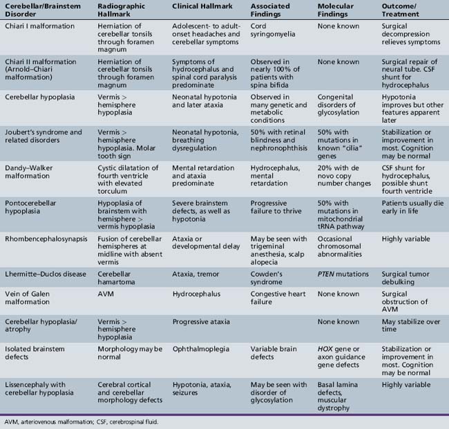
This table details various congenital and acquired cerebellar/brainstem disorders, outlining their key radiographic and clinical features. Conditions like Chiari and Dandy-Walker malformations are important to recognize.
Conclusion
In this lesson, we have dissected the cerebellum's role as the master coordinator of movement. We've moved from its basic anatomy to its functional organization and, most critically, to the clinical signs that arise when it is damaged.
Key Takeaways for Your Exam Revision:
- The cerebellum is a comparator, refining movement for accuracy and smoothness. It does not initiate movement.
- Lesions are ipsilateral, causing deficits on the same side of the body.
- Remember the DANISH mnemonic for the cardinal signs of cerebellar dysfunction: Dysdiadochokinesia, Ataxia, Nystagmus, Intention tremor, Slurred speech, Hypotonia.
- Midline lesions (vermis) cause truncal/gait ataxia.
- Hemispheric lesions cause ipsilateral limb (appendicular) ataxia.
- Be able to contrast cerebellar intention tremor with the resting tremor of Parkinson's disease.
Having covered the basal ganglia and the cerebellum, you now have a solid foundation for understanding the major subcortical motor systems. In our next lesson, we will begin our exploration of the cranial nerves, starting with nerves I through VI. These nerves provide the crucial sensory and motor links between the brain and the head, neck, and special sense organs.