Hello! In our last lesson, we navigated the critical topic of potassium abnormalities, focusing on how to recognize the life-threatening ECG patterns of hyperkalemia and the immediate need for cardiac membrane stabilization. That concept of an electrolyte's effect being dependent on its physiological state is a perfect bridge to today's topic.
Today, we will dissect the last major electrolyte on the Basic Metabolic Panel: calcium. Our goal is to interpret calcium abnormalities, correcting for albumin levels when necessary. A significant portion of calcium in the blood is bound to protein, which can create a misleading picture if not accounted for. Mastering this correction is a fundamental skill for accurately diagnosing and managing a wide range of common and critical conditions you'll encounter on the wards.
Calcium: More Than Just a Number
When you order a "calcium level" as part of a BMP, you typically get a total calcium measurement. However, calcium exists in the blood in three main forms:
- Ionized (or Free) Calcium: This is the physiologically active form that participates in neuromuscular signaling, cardiac contraction, and blood clotting. Its level is tightly regulated by the body.
- Protein-Bound Calcium: About 40-45% of total calcium is bound to proteins, primarily albumin. This portion is inactive and serves as a reservoir.
- Complexed Calcium: A small fraction is bound to anions like citrate and phosphate.
Because the ionized calcium is what truly matters for cellular function, but the total calcium is what's most commonly measured, we have a potential problem: what happens if the protein levels change?
To understand this crucial relationship, please watch the video below. It provides an excellent visual explanation of why a low albumin level can create a "false" hypocalcemia.
This video, 'Corrected Calcium Explained' by Dr. Mariam, clearly illustrates the relationship between total calcium, ionized calcium, and albumin.
Please watch from the beginning to 01:49. Focus on understanding: The difference between total, bound, and ionized calcium. Why a low albumin level causes the total calcium to drop while the ionized calcium can remain normal.
As the video explains, if a patient has low albumin (hypoalbuminemia), there are fewer binding sites for calcium. This reduces the protein-bound portion, causing the total calcium level to fall. However, the body's regulatory mechanisms (involving parathyroid hormone and vitamin D) work to keep the physiologically active ionized calcium in the normal range. Therefore, a low total calcium in a patient with low albumin might not represent a true calcium deficit at all. This is why we must correct for it.
The Correction Formula: Adjusting for Albumin
To estimate the "true" calcium level as if the albumin were normal, we use a standard correction formula. The general principle is that for every 1 g/dL decrease in serum albumin below a normal level of 4.0 g/dL, the total calcium level is expected to decrease by about 0.8 mg/dL.
The formula is:
Corrected Calcium (mg/dL) = Measured Total Calcium (mg/dL) + 0.8 * (4.0 - Serum Albumin [g/dL])
Let's watch the next segment of the video to see this formula in action.
Let's continue with the 'Corrected Calcium Explained' video to see a worked example of the formula.
Please watch from 01:49 to 03:27. The video walks through a clinical scenario, plugging the lab values into the formula to determine the corrected calcium.

Test your understanding!
You are reviewing labs for a 72-year-old patient admitted with poor nutritional intake. The BMP returns with the following values:
- Total Calcium: 7.9 mg/dL (Normal: 9.0-10.5 mg/dL)
- Albumin: 2.0 g/dL (Normal: 3.5-5.5 g/dL)
Does this patient have true hypocalcemia? Calculate the corrected calcium.
Show answer
Let's use the formula:
Corrected Calcium = Measured Total Calcium + 0.8 * (4.0 - Serum Albumin)
Corrected Calcium = 7.9 + 0.8 * (4.0 - 2.0)
Corrected Calcium = 7.9 + 0.8 * (2.0)
Corrected Calcium = 7.9 + 1.6
Corrected Calcium = 9.5 mg/dL
The corrected calcium level is 9.5 mg/dL, which is within the normal range. This patient has hypoalbuminemia causing a low total calcium, but does not have true hypocalcemia. Their ionized calcium level is likely normal.
The Gold Standard: Ionized Calcium and Clinical Nuances
While the correction formula is a widely used and tested tool, it's important to recognize its limitations.
The Medscape article 'Serum Calcium' provides an important clinical perspective on the reliability of correction formulas, especially in complex patients.
Please read the two short paragraphs under the 'Interpretation' and 'Background' headings that discuss the limitations of adjusted calcium calculations. Start with the sentence 'Traditional teaching advocates adjusting total calcium...' and end with '...direct measurement of ionized calcium is preferred over formulaic adjustment.' Then read the paragraph under 'Background' starting 'The clinical utility of "corrected" (albumin-adjusted) calcium...'
As the article points out, in certain populations—especially critically ill patients, those with kidney disease, or patients with acid-base disturbances—the formula can be inaccurate. In these cases, or whenever there is clinical doubt, the best course of action is to directly measure the ionized calcium.
An ionized calcium level does not need to be corrected for albumin, making it the "gold standard" measurement. However, as the first video mentioned, it's a more expensive test and the sample requires special handling, which is why total calcium remains the first-line screening test.
The Effect of pH
Another important factor that can affect calcium is the blood pH.
- Alkalosis (high pH) increases the negative charge on albumin, causing it to bind more calcium. This decreases the ionized calcium level, which can cause symptoms of hypocalcemia even if total calcium is normal.
- Acidosis (low pH) does the opposite, decreasing calcium binding and increasing the ionized calcium level.
This is particularly relevant in patients who are hyperventilating (causing respiratory alkalosis), who may develop tingling and muscle cramps (symptoms of hypocalcemia) due to this shift.
Interpreting Abnormal Calcium Levels
Once you have an accurate calcium value (either corrected or ionized), you can interpret it in the context of the patient's clinical picture.
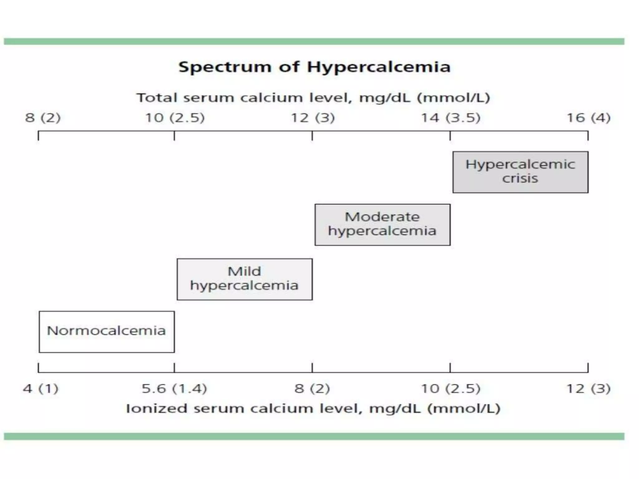
Hypocalcemia (Corrected Ca < 9.0 mg/dL)
- Key Symptoms: Due to increased neuromuscular excitability. Think "CATS go numb": Convulsions, Arrhythmias (prolonged QT interval), Tetany (involuntary muscle spasms, e.g., Chvostek's and Trousseau's signs), and numbness/paresthesias.
- Common Causes: Hypoparathyroidism (e.g., after neck surgery), severe Vitamin D deficiency, chronic kidney disease, and pancreatitis.
Hypercalcemia (Corrected Ca > 10.5 mg/dL)
- Key Symptoms: Due to decreased neuromuscular excitability. The classic mnemonic is "stones, bones, groans, and psychiatric overtones" (stones: kidney stones; bones: bone pain; groans: abdominal pain, constipation; psychiatric overtones: confusion, lethargy, depression). On an ECG, it can cause a short QT interval.
- Common Causes: Over 90% of cases are due to either primary hyperparathyroidism or malignancy.
To begin differentiating the causes, the next crucial step is to look at the calcium level in conjunction with the Parathyroid Hormone (PTH) level. This relationship is a critical diagnostic pattern.
Hypercalcemia and Hypocalcemia - Vitamin D Testing
This table from ARUP Consult provides a simplified but powerful framework for interpreting calcium and PTH levels together.
Please review the table titled 'Interpretation of Calcium and PTH Test Results'. Focus on the four main patterns: Low Ca, Low/Normal PTH Low Ca, High PTH High Ca, Low PTH High Ca, Normal/High PTH
This table demonstrates the core feedback loop:
- If calcium is low, PTH should be high (the body is trying to compensate). If it's not, the parathyroid gland itself is the problem (hypoparathyroidism).
- If calcium is high, PTH should be low (the body is trying to shut down production). If it's high or even "inappropriately normal," the parathyroid gland is likely overactive (hyperparathyroidism).
Conclusion
You've now learned the essential skill of interpreting serum calcium, a process that goes beyond simply looking at the reported number.
Key Takeaways:
- Ionized calcium is the physiologically active form, but total calcium is most often measured.
- Low serum albumin will artifactually lower the total calcium level.
- Always calculate a corrected calcium in patients with abnormal albumin using the formula:
Corrected Ca = Total Ca + 0.8 * (4.0 - Albumin). - In critically ill patients or when in doubt, measuring an ionized calcium level is the most accurate approach.
- The initial step in diagnosing a true calcium abnormality is to interpret it in the context of the PTH level.
In our next lesson, we will begin to synthesize what we've learned about individual electrolytes. We'll move from analyzing single values to interpreting the entire Basic Metabolic Panel to identify patterns of dehydration, a skill that requires integrating multiple data points to form a cohesive clinical picture.