Hello! In our last lesson, we worked through the systematic approach to classifying hyponatremia by assessing volume status and using key urine studies. We saw how this algorithm helps turn a simple lab value into a specific diagnosis.
Today, we continue our exploration of the Basic Metabolic Panel (BMP) by focusing on another critical electrolyte: potassium. Our goal is to interpret potassium abnormalities and recognize the ECG changes associated with life-threatening hyperkalemia. As a future physician, this is one of the most important lab-to-ECG correlations you will master, as prompt recognition can be life-saving. We'll cover the diagnostic framework for both low and high potassium and pay special attention to the urgent patterns that demand immediate action.
Potassium: The Basics of Balance
Potassium is the most abundant intracellular cation, and its concentration gradient across the cell membrane is crucial for the electrical activity of excitable tissues, especially cardiac and skeletal muscle. The normal serum potassium level is kept within a very narrow range, typically 3.5 to 5.2 mEq/L (or mmol/L).
- Hypokalemia is defined as a serum potassium < 3.5 mEq/L.
- Hyperkalemia is defined as a serum potassium > 5.2 mEq/L, with levels > 5.5 mEq/L often considered clinically significant and levels > 6.5 mEq/L becoming life-threatening.
To understand how to interpret abnormalities, we first need a framework for the causes. The video below provides an excellent overview.
Potassium Disorders | Clinical Medicine
The Ninja Nerd video 'Potassium Disorders' provides a fantastic conceptual framework for thinking about potassium abnormalities. We'll use short clips to establish the basic definitions and the major categories of causes.
Please watch three short segments: 00:00:52 - 00:01:14: Defines the numerical ranges for hypokalemia and hyperkalemia. 00:01:14 - 00:02:45: Introduces the three main causes of hypokalemia: intracellular shifting, GI loss, and renal loss. 00:38:17 - 00:40:27: Introduces the two main causes of hyperkalemia: intracellular shifting and renal retention.
As the video outlines, nearly all potassium abnormalities can be traced back to two fundamental processes:
- Transcellular Shifts: Potassium moving into or out of cells.
- Imbalance of Intake vs. Excretion: Primarily related to GI or renal losses (for hypokalemia) and renal retention (for hyperkalemia).
Hypokalemia: Causes and ECG Findings
While less acutely dangerous than severe hyperkalemia, hypokalemia is very common and can lead to significant morbidity, including dangerous tachyarrhythmias.
Common Causes of Hypokalemia:
- Renal Losses: Diuretic therapy (loop and thiazide diuretics) is a leading cause. Hyperaldosteronism also causes renal potassium wasting.
- GI Losses: Diarrhea and vomiting are classic causes of potassium and volume loss.
- Intracellular Shifts:
- Insulin: Drives potassium into cells. This is why patients being treated for diabetic ketoacidosis (DKA) can rapidly become hypokalemic.
- Beta-2 Agonists: Drugs like albuterol can also shift potassium into cells.
- Alkalosis: The body exchanges extracellular potassium for intracellular hydrogen ions to buffer the blood, lowering serum potassium.
Clinical Impact and ECG Changes in Hypokalemia
Hypokalemia makes nerve and muscle cells hyperpolarized, which paradoxically leads to cardiac hyperexcitability. This increases the risk for arrhythmias.
The classic ECG findings are a direct reflection of this altered repolarization.
Potassium Disorders | Clinical Medicine
Let's return to the Ninja Nerd video to understand the pathophysiology and see what these ECG changes look like.
Please watch from 01:03:23 to 01:04:45 and then from 01:17:15 to 01:20:23. The first part explains why cardiac excitability increases. The second part details the specific ECG findings you need to recognize.
Key ECG Findings in Hypokalemia:
- T-wave flattening or inversion
- ST-segment depression
- Prominent U-waves: A small wave appearing after the T wave, best seen in the precordial leads (V2-V4).
- Apparent long QT interval: Often due to the T-wave and U-wave merging (long QU interval). This state creates a risk for Torsades de Pointes, a life-threatening polymorphic ventricular tachycardia.
Hyperkalemia: A True Medical Emergency
Hyperkalemia is one of the most dangerous electrolyte abnormalities because it can lead to sudden cardiac arrest with minimal warning. You must be able to recognize its signature on an ECG.
Common Causes of Hyperkalemia:
- Decreased Renal Excretion: This is the most common cause.
- Acute or Chronic Kidney Disease (CKD): The kidneys simply cannot filter potassium. Patients who miss hemodialysis are at extremely high risk.
- Medications: ACE inhibitors, ARBs, potassium-sparing diuretics (e.g., spironolactone), and NSAIDs all impair the kidney's ability to excrete potassium.
- Intracellular Shifts (Potassium moving out of cells):
- Acidosis: The body buffers by shifting hydrogen ions into cells in exchange for potassium.
- Cell Lysis: Massive cell death releases large amounts of intracellular potassium. Think of rhabdomyolysis, tumor lysis syndrome, or major crush injuries.
- Insulin Deficiency (DKA): Lack of insulin impairs the Na⁺/K⁺ pump's ability to push potassium into cells.
Recognizing Life-Threatening Hyperkalemia on an ECG
Hyperkalemia decreases myocardial excitability and slows conduction through the heart. This creates a predictable, progressive sequence of ECG abnormalities as the potassium level rises.
Hyperkalemia EKG Findings - ECG Interpretation
The Medzcool video 'Hyperkalemia EKG Findings' provides a short and highly memorable overview of this progression.
Watch the entire video (00:00 - 03:34). Pay close attention to the 'pulling the string' analogy and the step-wise progression of changes.
This progression is one of the most important patterns to burn into your memory.
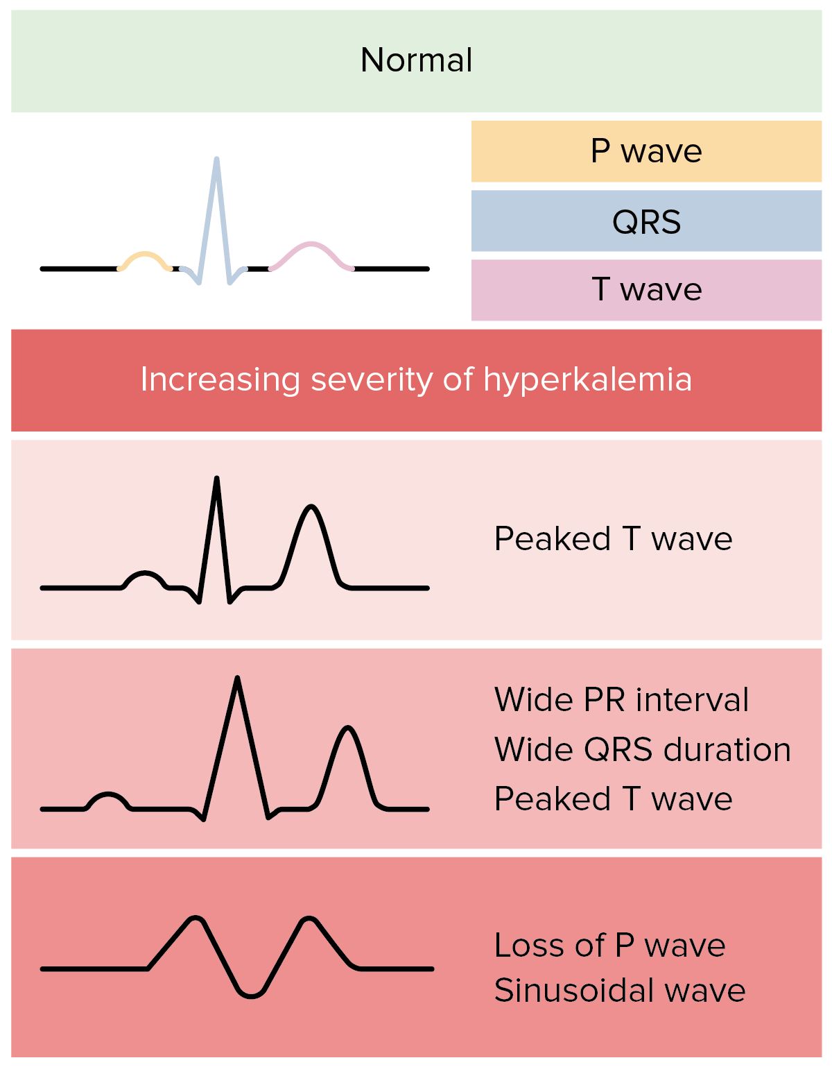
Let's walk through the stages:
- Mild Hyperkalemia (K⁺ > 5.5 mEq/L): Peaked T-waves. The T-waves become tall, narrow, and tent-like. This is the earliest sign.
- Moderate Hyperkalemia (K⁺ > 6.5 mEq/L): PR interval prolongation and P-wave flattening. The P-wave may shrink and eventually disappear as the atria fail to conduct.
- Severe Hyperkalemia (K⁺ > 7.0 mEq/L): QRS complex widening. The slowing of ventricular conduction causes the QRS to spread out.
- Very Severe/Pre-Arrest (K⁺ > 8.0 mEq/L): Sine Wave Morphology. The widened QRS merges with the T-wave, creating a smooth, undulating sine wave pattern. This is a harbinger of imminent cardiac arrest (ventricular fibrillation or asystole).
To solidify your pattern recognition, let's look at some real-world examples.
The ECG Library from 'Life in the Fastlane' (LITFL) is an exceptional resource for emergency medicine and ECG interpretation. It provides numerous real-world examples.
Please read through the webpage, paying special attention to the 'ECG Examples' section. Compare each ECG to the serum potassium level provided. Also, note the crucial clinical pearl: serum potassium may not correlate perfectly with ECG changes, and cardiac arrest can occur suddenly.
Test your understanding!
You are called to see a 68-year-old male with end-stage renal disease who missed his dialysis session. The nurse hands you this ECG.
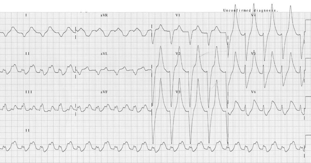
- Identify at least three abnormalities on this ECG.
- What is the most likely underlying electrolyte disturbance?
- Is this a medical emergency? What is the immediate physiological goal of treatment?
Show answer
- The ECG shows (1) marked QRS widening, (2) absent P-waves, and (3) tall, peaked T-waves. The overall morphology is approaching a sine wave.
- This is a classic ECG of severe hyperkalemia.
- Yes, this is an immediate life-threatening emergency. The immediate goal is not to lower potassium, but to stabilize the cardiac membrane to prevent arrhythmia and cardiac arrest.
The Critical First Step: Stabilize the Membrane
When you see ECG changes from hyperkalemia, the first and most important action is to administer intravenous calcium (calcium gluconate or calcium chloride).
Important: Calcium does NOT lower serum potassium. It works by antagonizing the toxic effect of potassium on the cardiac cell membrane, temporarily restoring normal excitability and preventing lethal arrhythmias. This buys you time to implement therapies that actually lower the potassium level (like insulin, albuterol, and dialysis).

Conclusion
You have now covered the essentials of interpreting potassium disorders, a high-yield topic for both exams and clinical practice.
Key Takeaways:
- Potassium abnormalities are caused by transcellular shifts or an imbalance in intake versus excretion (primarily renal).
- Hypokalemia can cause cardiac hyperexcitability, with ECG findings of T-wave flattening, ST depression, and prominent U-waves, increasing the risk of tachyarrhythmias.
- Hyperkalemia is a medical emergency that causes a predictable sequence of ECG changes: peaked T-waves → prolonged PR → widened QRS → sine wave → cardiac arrest.
- Recognizing these ECG changes is critical and the immediate response for an unstable membrane is IV calcium.
In our next lesson, we will move on to the final electrolyte in the BMP, calcium. We will learn to interpret calcium abnormalities, correcting for albumin levels when necessary, another crucial skill for managing hospitalized patients.