Hello! In our last lesson, we introduced the broad framework for interpreting sodium abnormalities. We established that sodium disorders are fundamentally problems of water balance and that the first steps in diagnosis involve checking serum osmolality and assessing the patient's volume status.
Today, we'll build directly on that foundation. Our goal is to master the classification of the most common type of hyponatremia—hypotonic hyponatremia—by using volume status and key urine studies. This systematic approach is essential for accurately narrowing down the wide range of possible causes you'll encounter in clinical practice.
Step 1: The First Gate – Confirming Hypotonic Hyponatremia
As we reviewed, before you can diagnose the cause of hyponatremia, you must first confirm you are dealing with hypotonic hyponatremia. This is "true" hyponatremia, where there is an excess of water relative to sodium.
Let's quickly refresh this crucial first step.
Your Easy Guide To Hyponatremia
The video 'Your Easy Guide To Hyponatremia' by Dr. Conan Liu provides a concise overview of the initial diagnostic questions. It clearly explains how to use serum osmolality to differentiate the types of hyponatremia.
Please watch from 00:34 to 02:59. Pay close attention to the three categories based on serum osmolality (hypotonic, isotonic, hypertonic) and how to calculate the corrected sodium in the setting of hyperglycemia.
To summarize the key points from the video:
- Measure Serum Osmolality (Sosm):
- Low Sosm (< 280 mOsm/kg): This is hypotonic hyponatremia. Proceed with the algorithm below.
- Normal Sosm (280-295 mOsm/kg): This suggests isotonic hyponatremia (or "pseudohyponatremia"), a lab artifact from severe hyperlipidemia or hyperproteinemia.
- High Sosm (> 295 mOsm/kg): This is hypertonic hyponatremia, most often caused by hyperglycemia, where glucose acts as an osmolyte, pulling water into the extracellular space and diluting the sodium.
Remember to calculate the corrected sodium for hyperglycemia:
Note: The correction factor can vary in literature (from 1.6 to 2.4), but 2.0 is a commonly used and practical estimate.
From this point forward, our lesson will focus exclusively on working up hypotonic hyponatremia.
Step 2: The Three Buckets – Classifying by Volume Status
Once you've confirmed hypotonic hyponatremia, the next step is a careful clinical assessment of the patient's volume status. This divides the potential causes into three main categories: hypovolemic, euvolemic, and hypervolemic. We then use urine studies—primarily urine sodium (UNa) and urine osmolality (Uosm)—to confirm our assessment and pinpoint the diagnosis.
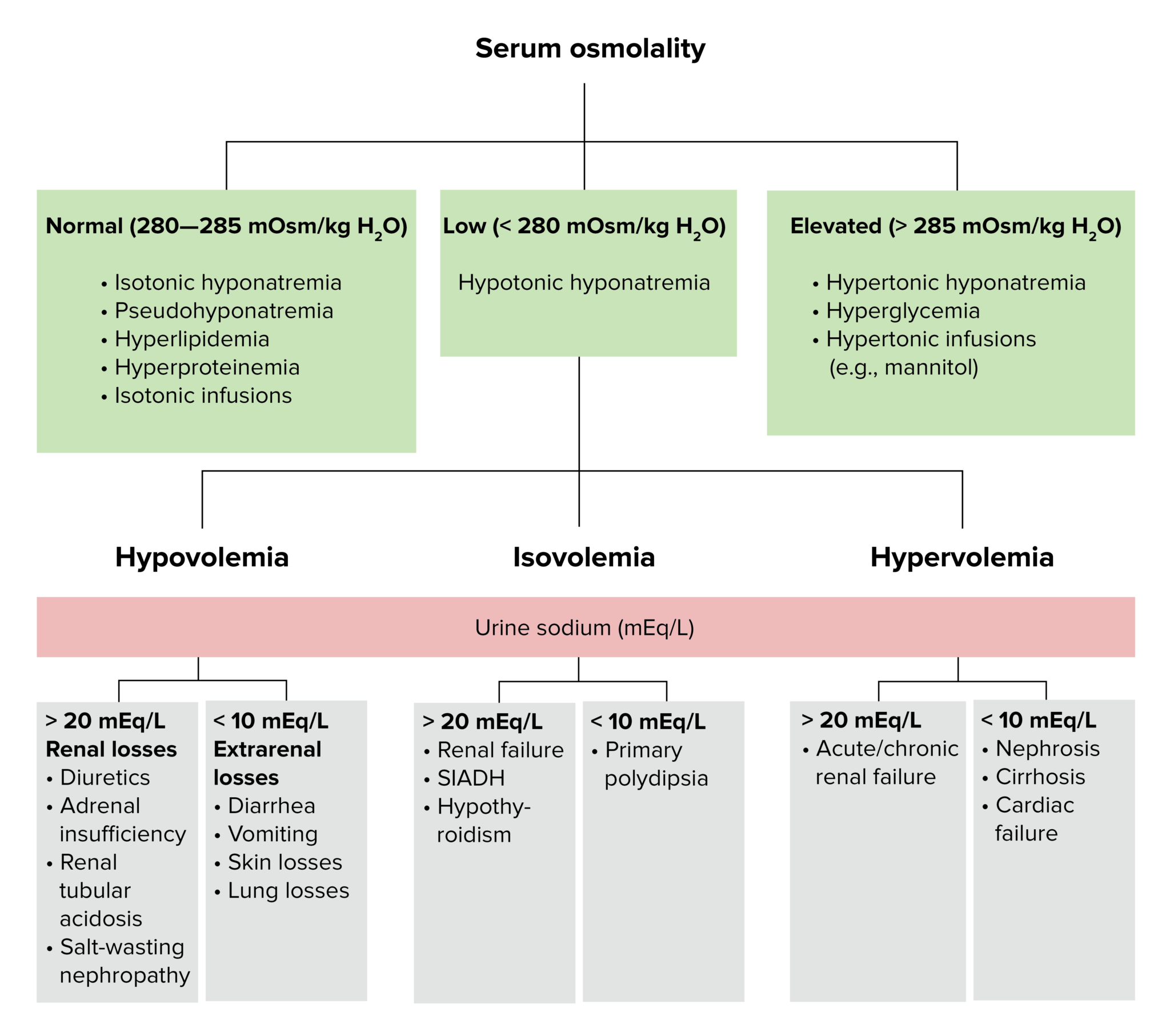
Let's break down each of these three "buckets."
A. Hypovolemic Hyponatremia (Dehydrated)
In this state, the patient has lost both salt and water, but the water loss is relatively less than the salt loss, or they have replaced losses with hypotonic fluids (like free water). The body's response to volume depletion is a powerful release of ADH to conserve water, which overrides the need to maintain normal osmolality.
To distinguish the cause of volume loss, we check the urine sodium:
- UNa < 20 mEq/L (Extra-renal losses): The kidneys are functioning correctly and are trying to conserve sodium in response to volume depletion. This points to losses from outside the kidneys.
- Common Causes: GI losses (vomiting, diarrhea), third-spacing (burns, pancreatitis, bowel obstruction), excessive sweating.
- UNa > 20 mEq/L (Renal losses): The kidneys are the source of the salt and water loss. They are inappropriately excreting sodium despite volume depletion.
- Common Causes: Diuretic use (especially thiazides), primary adrenal insufficiency (Addison's disease), salt-losing nephropathies.
Diagnosis and Management of Sodium Disorders
The AAFP article 'Diagnosis and Management of Sodium Disorders' provides a useful table summarizing the causes and diagnostic clues for hypovolemic hyponatremia.
Please read the section titled 'HYPOVOLEMIC HYPONATREMIA' and review the accompanying table. Notice how the clinical context combined with the urine sodium level helps distinguish between causes like diuretic use and GI loss.
B. Hypervolemic Hyponatremia (Edematous / Fluid Overloaded)
Here, the patient has an excess of both total body sodium and water, but the water gain is greater. This leads to clinical signs of fluid overload like peripheral edema, pulmonary edema (rales), and ascites. ADH is elevated because these conditions (heart failure, cirrhosis) lead to a low effective circulating volume, even though total body volume is high. The body perceives under-perfusion and tries to retain water.
Again, we use urine sodium to differentiate the cause:
- UNa < 20 mEq/L (Intact renal perfusion sensing): The kidneys sense low effective volume and avidly reabsorb sodium.
- Common Causes: Congestive heart failure (CHF), Cirrhosis, Nephrotic syndrome.
- UNa > 20 mEq/L (Kidney failure): The kidneys themselves are failing and can no longer effectively reabsorb sodium.
- Common Causes: Acute or chronic renal failure.
C. Euvolemic Hyponatremia (Clinically Normal Volume)
This is often the most challenging category. These patients have a modest excess of total body water, but not enough to be clinically evident as edema. The key feature is the inappropriate secretion of ADH, unrelated to either osmolarity or volume status.
Here, we use both urine osmolality and urine sodium:
- Uosm > 100 mOsm/kg: This indicates that ADH is active, causing the kidneys to produce concentrated urine. This is inappropriate in a patient who is already hypotonic.
- UNa > 20-40 mEq/L: Since the patient is not volume-depleted, the renin-angiotensin-aldosterone system (RAAS) is not activated, and the kidneys continue to excrete sodium taken in through the diet.
The combination of euvolemia, hypotonicity, high Uosm, and high UNa is the classic profile for Syndrome of Inappropriate ADH (SIADH).
Differential Diagnosis for Euvolemic Hyponatremia:
- SIADH: This is a diagnosis of exclusion. Always consider its common causes:
- CNS disorders (stroke, hemorrhage, infection)
- Pulmonary diseases (pneumonia, TB, cancers)
- Malignancy (especially small cell lung cancer)
- Drugs (SSRIs, carbamazepine, opioids, ecstasy)
- Pain, nausea, post-operative state
- Hypothyroidism: Severe hypothyroidism can cause a picture similar to SIADH.
- Adrenal Insufficiency (Glucocorticoid deficiency): Cortisol normally suppresses ADH, so its absence leads to elevated ADH.
A Note on Clinical Nuance
Assessing volume status at the bedside can be surprisingly difficult. A patient who appears euvolemic might actually be subtly hypovolemic. This is where lab data becomes even more crucial.
Diagnosing Hyponatremia: A Comprehensive Algorithm
The Medmastery video 'Diagnosing Hyponatremia' highlights this clinical challenge and introduces another helpful lab test, serum uric acid, as a potential tie-breaker.
Please watch from 02:58 to 05:45. Focus on the discussion about the difficulty of assessing volume status and how urine sodium and serum uric acid can help biochemically differentiate between hypovolemic and euvolemic states. Note the important caveats about diuretic use.
As the video explains, in SIADH (euvolemic), uric acid is often low due to urinary wasting, while in hypovolemia, it's often high due to renal retention. This can be a useful clue when the clinical picture is ambiguous.
Test your understanding!
You are evaluating a 65-year-old man admitted for pneumonia who develops a sodium of 124 mEq/L. His mucous membranes are moist, and he has no edema.
Labs:
- Serum Na⁺: 124 mEq/L
- Serum Osmolality: 260 mOsm/kg
- Urine Osmolality: 450 mOsm/kg
- Urine Sodium: 55 mEq/L
- Is this hypotonic, isotonic, or hypertonic hyponatremia?
- What is his likely volume status based on the history and physical exam?
- Based on the urine studies, which of the three "buckets" does he fit into?
- What is the most likely diagnosis?
Show answer
- Hypotonic hyponatremia. The serum osmolality is low (< 280 mOsm/kg).
- Euvolemic. His exam shows no signs of dehydration or fluid overload.
- Euvolemic hyponatremia bucket. He has concentrated urine (Uosm > 100) and a high urine sodium (UNa > 40) in the setting of clinical euvolemia and hypotonicity.
- The most likely diagnosis is SIADH, likely caused by his underlying pneumonia.
Conclusion
Mastering the differential diagnosis of hyponatremia is a core skill in internal medicine. By applying this systematic approach, you can move from a confusing lab value to a focused and accurate diagnosis.
Key Takeaways:
- Always start by confirming hypotonic hyponatremia with a serum osmolality.
- Clinically assess the patient's volume status to place them into one of three buckets: hypovolemic, hypervolemic, or euvolemic.
- Use urine osmolality to confirm ADH activity and urine sodium to differentiate between renal and extra-renal causes or between volume-depleted and euvolemic states.
- The classic pattern for SIADH is euvolemia with an inappropriately high urine osmolality (>100) and urine sodium (>40).
In our next lesson, we will continue our exploration of the Basic Metabolic Panel by turning our attention to potassium. We will learn how to interpret potassium abnormalities and recognize the critical ECG changes associated with life-threatening hyperkalemia.