Hello! Welcome to the next module of our course. We've spent a lot of time mastering the complete blood count (CBC). Now, we're shifting gears from hematology to clinical chemistry and one of the most fundamental lab panels you'll order: the Basic Metabolic Panel (BMP), or "Chem-7".
Today, we'll tackle the first and arguably most complex component of the BMP. Our goal is to recognize and interpret sodium abnormalities (hyponatremia and hypernatremia). As you've likely seen on your rotations, sodium disorders are incredibly common and can signal anything from simple dehydration to a life-threatening endocrine or neurological emergency. Mastering a systematic approach to them is a vital clinical skill.
Part 1: Sodium, Osmolality, and Water Balance
Sodium is the most abundant cation in the extracellular fluid and the primary driver of plasma osmolality. This means it dictates where water goes. A change in sodium concentration reflects a disorder of water balance, not necessarily a disorder of total body sodium. This is the single most important concept to grasp.
When extracellular sodium concentration changes, water moves across cell membranes to maintain osmotic equilibrium.
- Hyponatremia (low sodium): Water moves into cells, causing them to swell. In the brain, this leads to cerebral edema and neurological symptoms.
- Hypernatremia (high sodium): Water moves out of cells, causing them to shrink. This leads to cellular dehydration, which is also particularly dangerous for brain cells.
The body defends plasma osmolality primarily through thirst and the action of antidiuretic hormone (ADH), which promotes water reabsorption in the kidneys.
To start, let's watch a brief segment from the 'Understanding Hyponatraemia' video by Zero To Finals. It provides a clear, concise review of osmolality and how it governs fluid movement.
Please watch from 01:07 to 03:26. Focus on understanding the relationship between plasma osmolality (driven by sodium) and the movement of water between the intracellular and extracellular compartments.
Part 2: Hyponatremia (Serum Na⁺ < 135 mEq/L)
Hyponatremia is the most common electrolyte abnormality in hospitalized patients. Symptoms depend on the severity and acuity, ranging from mild nausea and malaise to severe confusion, seizures, and coma.
Interpreting hyponatremia requires a structured, three-step approach.
Step 1: Check Serum Osmolality
The first step is to determine if you are dealing with a true, hypotonic hyponatremia.
- Hypotonic Hyponatremia (Serum Osmolality < 280 mOsm/kg): This is "true" hyponatremia, where there is an excess of water relative to solute. This is the most common type and will be our focus.
- Isotonic Hyponatremia (Serum Osmolality 280-295 mOsm/kg): This is a lab artifact, often called "pseudohyponatremia." It occurs with very high levels of lipids or proteins, which reduce the percentage of water in the plasma sample. The sodium concentration in the water portion is actually normal.
- Hypertonic Hyponatremia (Serum Osmolality > 295 mOsm/kg): This occurs when another osmotically active substance (most commonly glucose in hyperglycemia) pulls water into the extracellular space, diluting the sodium. You must always correct the measured sodium for hyperglycemia.
Diagnosis and Management of Sodium Disorders
The article 'Diagnosis and Management of Sodium Disorders' from American Family Physician provides a good summary of the initial tests.
Please read the 'LABORATORY TESTING' section. Pay attention to the role of serum osmolality in differentiating the types of hyponatremia. Note the table that lists initial tests and their considerations.
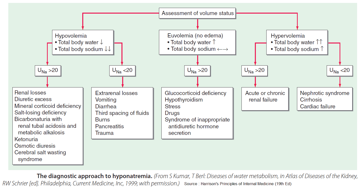
Step 2 & 3: Assess Volume Status and Check Urine Studies
Once you've confirmed hypotonic hyponatremia, the next crucial step is to assess the patient's volume status, which narrows the differential diagnosis considerably. We then use urine sodium (UNa) to distinguish between renal and extrarenal causes.
The following video provides an excellent walkthrough of this entire diagnostic algorithm.
Let's return to the Zero To Finals video. This extended segment walks through the full clinical algorithm for diagnosing the cause of hyponatremia.
Please watch from 07:13 to 12:16. Follow along as the video explains how to use volume status (dehydrated, euvolemic, edematous) and urine studies (urine sodium/osmolality) to pinpoint the diagnosis.
The diagnostic approach can be summarized by the following algorithm:
A Note on a Critical Complication: ODS
Rapid correction of chronic hyponatremia (> 48 hours) can lead to a devastating neurological condition called Osmotic Demyelination Syndrome (ODS), formerly central pontine myelinolysis. As the brain adapts to chronic low sodium, a sudden increase in extracellular sodium pulls water out of brain cells too quickly, causing myelin sheath damage.
To prevent this, the correction rate should be limited.
- Correction Goal: Increase serum sodium by no more than 8-10 mEq/L in the first 24 hours and no more than 18 mEq/L in the first 48 hours.
Part 3: Hypernatremia (Serum Na⁺ > 145 mEq/L)
Hypernatremia is almost always a free water deficit. The patient has lost water, is not drinking enough water, or, rarely, has ingested too much salt. It is always a hypertonic state.
Symptoms are primarily neurological and due to brain cell shrinkage, including lethargy, weakness, and, if severe, seizures and coma. The diagnostic approach is also based on volume status.
Hypernatremia Explained Clearly - Pathophysiology & Treatment
Now let's turn to hypernatremia. The MedCram video 'Hypernatremia Explained Clearly' provides a fantastic breakdown of the pathophysiology and classification based on volume status.
Please watch from the beginning to 15:42. The video systematically explains hypovolemic, hypervolemic, and isovolemic hypernatremia, linking each to its specific causes and pathophysiology.
Here is a summary of the approach to hypernatremia:

Key causes to remember:
- Hypovolemic Hypernatremia: Dehydration from GI losses, sweating, or diuretics, especially in patients with an impaired thirst mechanism or lack of access to water (e.g., elderly, infants, altered mental status). This is the most common cause.
- Euvolemic Hypernatremia: Pure water loss. This is the hallmark of Diabetes Insipidus (DI), which can be central (no ADH production) or nephrogenic (kidneys don't respond to ADH).
- Hypervolemic Hypernatremia: Gain of hypertonic fluid. This is almost always iatrogenic, from the administration of hypertonic saline or sodium bicarbonate.
Test your understanding!
Case 1: An 82-year-old woman from a nursing home is brought to the ED with confusion. She has dry mucous membranes and poor skin turgor.
- Labs: Serum Na⁺ 128 mEq/L, Serum Osm 265 mOsm/kg, Urine Na⁺ 15 mEq/L.
- What is the type of hyponatremia and the most likely cause?
Case 2: A 35-year-old patient with a history of severe bipolar disorder treated with lithium presents with extreme thirst and is urinating large volumes of dilute urine.
- Labs: Serum Na⁺ 152 mEq/L, Serum Osm 315 mOsm/kg, Urine Osm 150 mOsm/kg.
- What is the type of hypernatremia and the likely diagnosis?
Show answer
Case 1: This is hypovolemic hypotonic hyponatremia. The low serum osmolality confirms true hyponatremia. The clinical signs point to dehydration (hypovolemia). The low urine sodium (< 20 mEq/L) indicates the kidneys are appropriately conserving sodium, meaning the loss is extra-renal. The likely cause is dehydration due to poor oral intake.
Case 2: This is euvolemic hypernatremia. The high sodium and serum osmolality with inappropriately dilute urine (low urine osmolality) is the classic picture of Diabetes Insipidus (DI). Given the history of lithium use, which is a known cause of nephrogenic DI, the likely diagnosis is lithium-induced nephrogenic diabetes insipidus.
Conclusion
You have now learned the fundamental principles for interpreting sodium abnormalities. This framework will serve you well on the wards and in exams.
Key Takeaways:
- Sodium disorders are primarily disorders of water balance.
- The first step in evaluating hyponatremia is to check serum osmolality to confirm true hypotonicity.
- The cornerstone of diagnosis for both hyponatremia and hypernatremia is assessing the patient's volume status (hypovolemic, euvolemic, or hypervolemic).
- Urine sodium is critical for differentiating renal from extra-renal causes in hypovolemic states.
- Always be mindful of the safe rate of correction for chronic hyponatremia to prevent Osmotic Demyelination Syndrome.
In this lesson, we introduced the complete diagnostic algorithm for hyponatremia. In our next lesson, we will take a deeper dive, focusing specifically on how to classify hyponatremia by volume status and urine studies to narrow the differential diagnosis, reinforcing and expanding on the framework you learned today.