Hello! Welcome back.
In our last lesson, we focused on abnormalities in a single cell line—platelets—learning to spot urgent scenarios like TTP and DIC from a low count, or clonal disorders from a high one. We're now ready to zoom out and look at the "big picture" of the CBC. Sometimes, the most alarming patterns aren't just in one cell line, but across all of them, pointing to a central problem in the bone marrow, the factory where all blood cells are made.
Today's learning outcome is to recognize CBC patterns suggestive of bone marrow disorders, such as pancytopenia or the presence of blasts. This is a critical skill, as these patterns are often the first sign of life-threatening conditions like aplastic anemia or acute leukemia.
Part 1: Pancytopenia - When the Factory Shuts Down
Pancytopenia is a global failure of blood cell production, resulting in low counts of red cells, white cells, and platelets. As a clinician, when you see pancytopenia, your first thought should be: is this a problem with production in the bone marrow, or are the cells being destroyed in the periphery?
Approach to pancytopenia: From blood tests to the bedside
Let's start with a formal definition and a high-level overview from the article 'Approach to pancytopenia: From blood tests to the bedside'.
Please read the 'Introduction' section. Focus on the definition of pancytopenia and the broad categorization of causes into 'peripheral cell destruction' (like hypersplenism) and 'impaired bone marrow production' (like aplastic anemia or leukemia).
As you read, the core framework is production vs. destruction.
- Impaired Production: The bone marrow isn't making enough cells. This is the most common reason for severe pancytopenia.
- Peripheral Destruction: The marrow is working, but cells are being destroyed or sequestered elsewhere (e.g., an enlarged spleen).
Visually, severe bone marrow failure can be striking. Compare a normal, cellular marrow to one that has become fatty and empty in aplastic anemia.
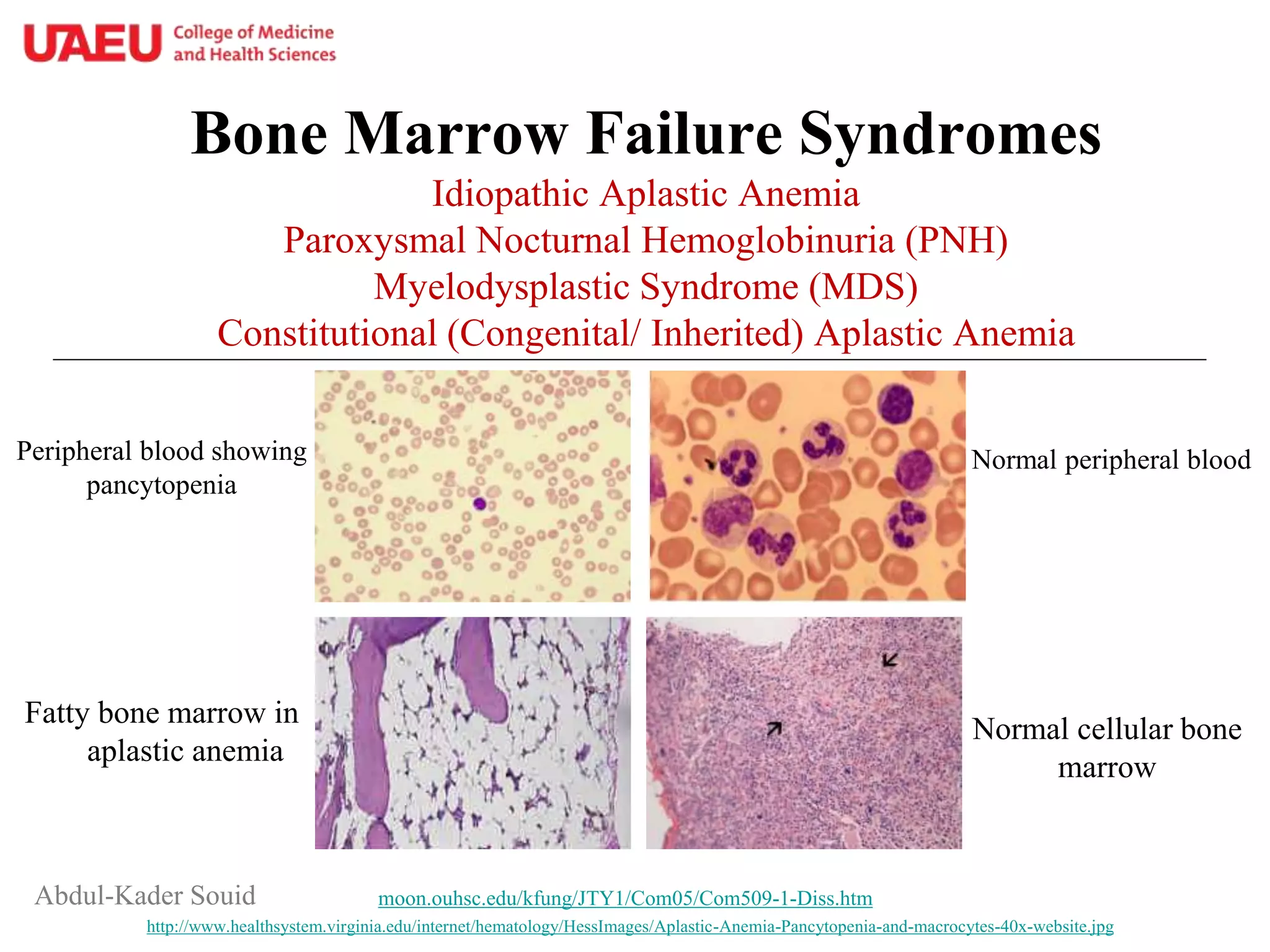
A Clinical Approach to Pancytopenia
The diagnostic journey for pancytopenia is a classic example of clinical detective work. It begins not with a complex test, but with a thorough history and physical exam. The video below, by a professor of medicine, provides an excellent case-based walkthrough of this process.
Approach to Pancytopenia by Dr Sudha Vidyasagar (Professor, Medicine)
Dr. Sudha Vidyasagar's 'Approach to Pancytopenia' provides a masterclass in clinical reasoning. We'll watch the introduction to establish the core principles.
Please watch from the beginning to 02:42 and then from 10:22 to 11:23. Pay close attention to two key points: The fundamental framework: Decreased production vs. Increased destruction. The single most important historical question: Is the pancytopenia acute or chronic? This one question dramatically changes your differential diagnosis.
Recognizing Patterns of Bone Marrow Failure
With that framework in mind, let's explore the patterns of specific bone marrow disorders that present with pancytopenia. Dr. Vidyasagar's lecture walks through several classic case presentations. As you review these summaries (based on the video), focus on the key features that differentiate one diagnosis from another.
-
Aplastic Anemia:
- The Pattern: Chronic pancytopenia with no other findings. No enlarged spleen or lymph nodes. The patient's only problem is the consequence of low blood counts (fatigue, infections, bleeding).
- The Cause: The bone marrow simply fails and is replaced by fat. It's often idiopathic.
- (Corresponds to video section 3, 11:59-16:04)
-
Myelodysplastic Syndrome (MDS):
- The Pattern: Pancytopenia in an older patient, sometimes with a mildly enlarged spleen. The key finding on the peripheral smear is dysplasia—abnormally shaped cells.
- The Cause: A pre-leukemic clonal disorder where the marrow is often cellular but produces defective, non-functional cells. It carries a risk of transforming into acute leukemia.
- (Corresponds to video section 4, 20:58-24:30)
-
Myelofibrosis:
- The Pattern: An older patient with constitutional symptoms (weakness, weight loss), pancytopenia, and a massively enlarged spleen. The peripheral smear is classic: teardrop-shaped red cells and a leucoerythroblastic picture (immature red and white cells spilling out of an irritated marrow).
- The Cause: The bone marrow is replaced by fibrous scar tissue.
- (Corresponds to video section 6, 27:07-29:46)
-
Marrow Infiltration (Metastases, Lymphoma):
- The Pattern: Pancytopenia in a patient with a known malignancy or with constitutional symptoms like weight loss. You may find lymphadenopathy (lymphoma) or clues to a primary tumor (e.g., urinary symptoms in prostate cancer).
- The Cause: Cancer cells invade the bone marrow, crowding out normal production.
- (Corresponds to video sections 8 & 9, 32:31-35:59)
Part 2: Blasts - When the Factory is Invaded
While pancytopenia can signify an empty or failing factory, the presence of blasts in the peripheral blood signifies something more sinister: the factory has been taken over by rogue, immature cells.
What is a blast? Blasts are the earliest precursors of blood cells. They are normally confined to the bone marrow in very small numbers (<5%). Morphologically, they are large cells with a high nuclear-to-cytoplasmic ratio, fine "open" chromatin, and prominent nucleoli.
Their appearance in the peripheral blood is a major red flag for acute leukemia.
This document from the International Clinical Cytometry Society provides a technical definition of blasts. We'll focus on the introduction.
Please read the 'Introduction' and 'Morphological features of blasts' sections (first page). Note the description of blasts as 'stem cell-derived, lineage-specific precursors' and the visual features that distinguish them from mature cells. Pay special attention to the mention of Auer rods, which are highly specific for acute myeloid leukemia (AML).
In acute leukemia, these malignant blasts lose their ability to mature but retain their ability to replicate endlessly. They proliferate and replace the normal bone marrow, leading to a catastrophic failure of hematopoiesis.
Acute Leukemia - ALL and AML - Hematology and Oncology Series
This video clearly explains how the proliferation of blasts leads to the clinical signs of acute leukemia.
Please watch the following segments: 03:47 - 05:14: Explains how blasts 'replace most of the bone marrow, crowding out normal hematopoiesis' and causing pancytopenia. 06:31 - 08:00: Describes the clinical features that result from this pancytopenia (fatigue from anemia, infections from neutropenia, bleeding from thrombocytopenia). 12:22 - 12:59: Emphasizes the key diagnostic criterion: more than 20% blasts in the bone marrow defines acute leukemia.
Part 3: Putting It All Together - Key CBC Patterns
You are now equipped to recognize two major patterns of bone marrow disorders on a CBC and peripheral smear report.
| CBC & Smear Pattern | Most Likely Diagnosis | Clinical Implication |
|---|---|---|
| Pancytopenia without blasts, low reticulocytes | Bone Marrow Failure (Aplastic Anemia, MDS, Infiltration) | Urgent, but diagnostic pathway involves history (acute vs. chronic) and physical exam (spleen, lymph nodes). |
| Any cytopenia(s) with circulating blasts present | Acute Leukemia | Hematologic Emergency. Requires immediate hematology consultation and likely a bone marrow biopsy. |
| Pancytopenia with teardrop cells and a leucoerythroblastic picture | Myelofibrosis (or other marrow infiltrative process) | Suggests a space-occupying lesion in the marrow. Often associated with massive splenomegaly. |
| Pancytopenia with dysplastic features (e.g., bilobed neutrophils, macro-ovalocytes) | Myelodysplastic Syndrome (MDS) | A "pre-leukemic" state that requires monitoring and supportive care. |
Test your understanding!
A 24-year-old presents to the ED with a week of high fevers, bleeding gums, and profound fatigue.
Her CBC shows:
- Hemoglobin: 7.1 g/dL (Low)
- WBC: 1.2 x 10⁹/L (Low)
- Platelets: 18,000/µL (Low)
- Peripheral smear comment: "Pancytopenia. 70% of white cells appear to be blasts. Auer rods noted."
What is the most likely diagnosis, and what is the significance of this pattern?
Show answer
The most likely diagnosis is Acute Myeloid Leukemia (AML). The combination of pancytopenia and a large percentage of circulating blasts is the classic pattern for acute leukemia. The presence of Auer rods is pathognomonic for AML. This is a hematologic emergency requiring immediate hospitalization and hematology consultation.
Conclusion
Today we've learned how to look at the CBC not just as a list of numbers, but as a window into the health of the bone marrow. You can now recognize the patterns that signal the factory is either shutting down or has been invaded.
Key Takeaways:
- Pancytopenia signals a global failure of hematopoiesis. Your first step is to determine if it's a production problem (aplastic anemia, MDS, infiltration) or a destruction problem (hypersplenism).
- A careful history (acute vs. chronic) and physical exam (looking for splenomegaly or lymphadenopathy) are essential to narrow the differential for pancytopenia.
- The presence of circulating blasts is a critical finding, highly suggestive of acute leukemia, a medical emergency. The formal diagnosis requires >20% blasts on a bone marrow biopsy.
Next Steps
We've now completed our deep dive into hematology and the CBC. In the next module, we will shift gears to clinical chemistry, starting with one of the most frequently ordered lab panels: the Basic Metabolic Panel (BMP). Our first lesson will focus on interpreting abnormalities in sodium.