Hello! Welcome back to our course on clinical lab interpretation.
In our last few lessons, we've systematically worked our way through the white blood cell differential. Today, we'll shift our focus to the final major component of the Complete Blood Count (CBC): the platelets.
Our learning outcome for this lesson is to identify thrombocytopenia and thrombocytosis and recognize when urgent evaluation is needed. Platelet disorders can range from incidental and benign findings to true hematologic emergencies. Your ability to quickly distinguish between these scenarios is a critical clinical skill. We'll focus on the high-yield patterns you'll encounter on your rotations.
Part 1: Thrombocytopenia (Too Few Platelets)
Thrombocytopenia is defined as a platelet count below the normal range, typically < 150,000/µL (or 150 x 10⁹/L). The most immediate concern with a low platelet count is the risk of bleeding.
When is Thrombocytopenia an Emergency?
The urgency of evaluation depends on two key factors: the absolute platelet count and the clinical context. The article below provides an excellent framework for assessing bleeding risk.
Thrombocytopenia: Evaluation and Management
This article from the American Academy of Family Physicians, 'Thrombocytopenia: Evaluation and Management,' outlines the clinical approach to low platelets.
Please read the following sections: Start from the beginning and read down to the end of the 'Bleeding Risk' section. Pay close attention to the platelet count thresholds and their associated symptoms. Next, read the 'Inpatient Evaluation' section to understand the 'red flag' findings that mandate immediate hospitalization.
As you just read, the bleeding risk is not linear. Here's a practical summary of the key thresholds:
- Platelet Count > 50,000/µL: Patients are generally asymptomatic.
- Platelet Count 20,000 - 50,000/µL: May see signs of minor bleeding like petechiae (pinpoint red dots), purpura (larger purple spots), or easy bruising.
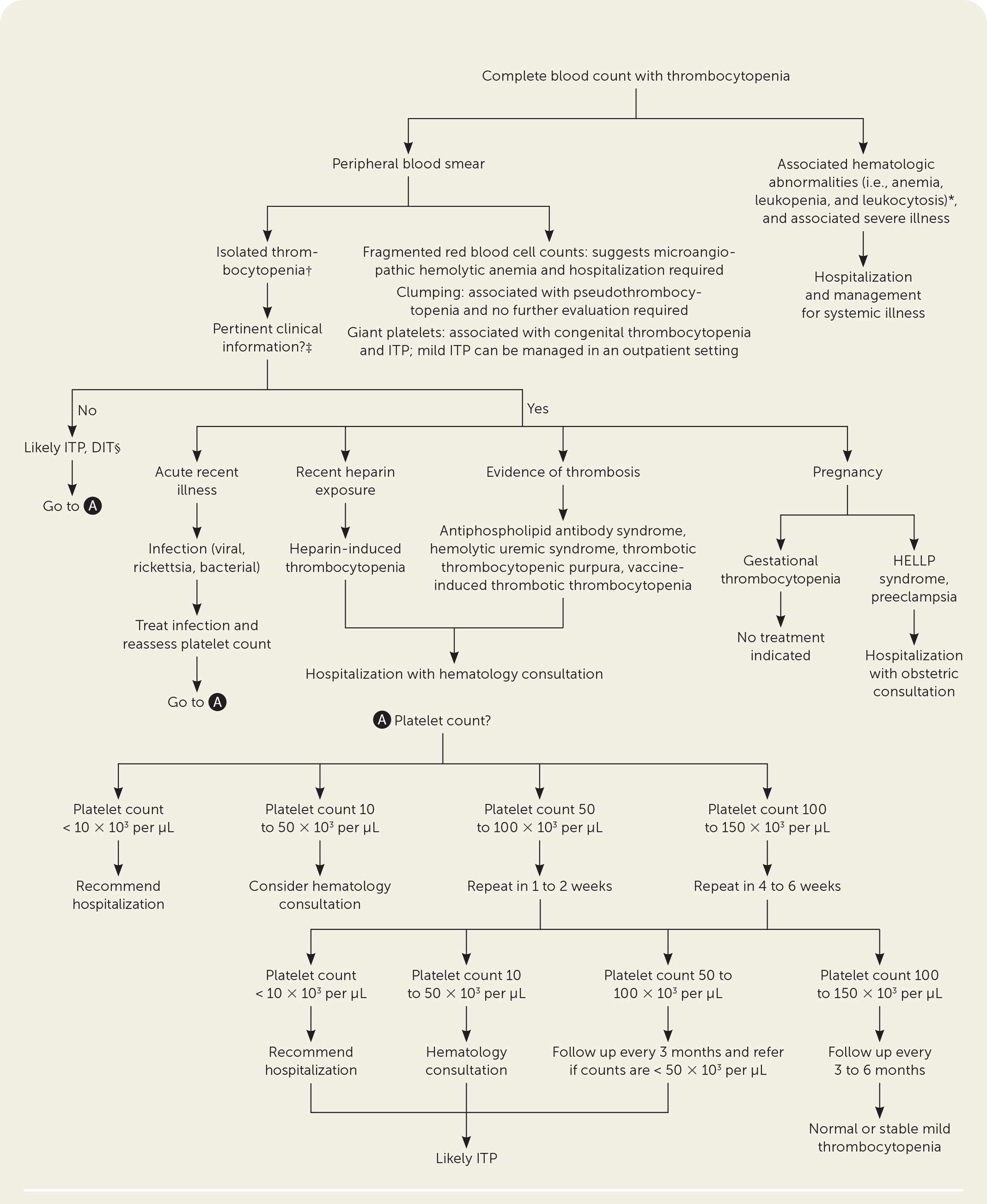
- Platelet Count < 10,000/µL: High risk of spontaneous, life-threatening hemorrhage (e.g., intracranial or GI bleeding). This is a hematologic emergency.
Any major bleeding or a platelet count <10,000/µL requires immediate action. Other red flags include new neurologic deficits, evidence of widespread clotting (thrombosis), or signs of red cell destruction (hemolysis).
The diagnostic algorithm below provides a great visual summary of how to approach a low platelet count.
Recognizing Emergency Patterns: TTP, HUS, DIC, and HIT
While many conditions can cause thrombocytopenia, there are a handful of life-threatening syndromes you must be able to recognize immediately. They often involve a paradoxical combination of low platelets and clotting.
The following video provides an outstanding, practical review of these critical conditions using mnemonics and clear explanations, which is perfect for building the pattern recognition skills you're aiming for.
Thrombocytopenia Review (ITP, TTP, HUS, DIC, HIT) | Mnemonics And Proven Ways To Memorize
This 'Cram The PANCE' video is a high-yield review of the most urgent causes of thrombocytopenia. It clearly explains the pathophysiology, clinical presentation, lab findings, and management of each.
Please watch from 00:19 to 34:23. This covers TTP, DIC, HUS, ITP, and HIT, followed by a helpful side-by-side comparison. As you watch, focus on identifying the unique 'signature' of each condition: TTP: The 'FAT RN' pentad and normal coags. DIC: The 'sick and bleeding' patient with abnormal coags. HUS: The child with bloody diarrhea and kidney failure. HIT: The patient on heparin who develops clots. ITP: The patient with isolated low platelets and no other abnormalities.
Let's summarize the key patterns from the video that signal an emergency:
| Condition | Key Clinical Clue | Key Lab Pattern | Urgent Action |
|---|---|---|---|
| TTP (Thrombotic Thrombocytopenic Purpura) | "FAT RN": Fever, Anemia, Thrombocytopenia, Renal dysfunction, Neurologic changes. | Schistocytes on smear, Normal PT/PTT. | Emergency plasmapheresis. Do not give platelets. |
| DIC (Disseminated Intravascular Coagulation) | "Sick and bleeding" patient (sepsis, trauma, malignancy). Oozing from IV sites. | Schistocytes, Elevated PT/PTT, Low Fibrinogen, High D-dimer. | Treat underlying cause, supportive care. |
| HUS (Hemolytic Uremic Syndrome) | Child with recent bloody diarrhea, acute kidney injury. | Schistocytes, Normal PT/PTT, Elevated Creatinine. | Supportive care. Avoid antibiotics. |
| HIT (Heparin-Induced Thrombocytopenia) | Patient on heparin develops thrombosis. >50% drop in platelet count. | Platelet drop 5-10 days after heparin start. Positive HIT antibodies. | Stop heparin immediately. Start a non-heparin anticoagulant. |
Immune Thrombocytopenic Purpura (ITP), also covered in the video, is a diagnosis of exclusion. It's characterized by isolated thrombocytopenia without the other features seen above (no schistocytes, normal coags). It becomes an emergency only when the platelet count is extremely low (<10-20k) or there is active, major bleeding.
Test your understanding!
A 58-year-old man is in the ICU with severe pneumonia and sepsis. You notice he has new bleeding from his IV lines and his Foley catheter. His labs show:
- Platelets: 45,000/µL
- PT/INR: 2.1 (elevated)
- PTT: 65 seconds (elevated)
- Fibrinogen: 80 mg/dL (low)
- D-dimer: 15,000 ng/mL (very high)
- Peripheral smear: Schistocytes noted.
What is the most likely diagnosis?
Show answer
The diagnosis is Disseminated Intravascular Coagulation (DIC). The combination of a critically ill ("sick") patient, new bleeding, severe thrombocytopenia, widespread activation of the clotting cascade (prolonged PT/PTT, high D-dimer), consumption of clotting factors (low fibrinogen), and RBC fragmentation (schistocytes) is the classic pattern for DIC.
Part 2: Thrombocytosis (Too Many Platelets)
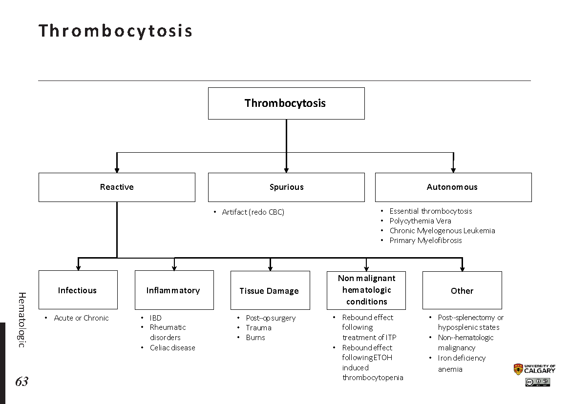
Thrombocytosis is an elevated platelet count, typically defined as > 450,000/µL. Unlike thrombocytopenia, most cases of thrombocytosis are not dangerous. The key is to distinguish a benign reactive process from a more serious clonal disorder.
Reactive vs. Clonal Thrombocytosis
-
Reactive (Secondary) Thrombocytosis: This is by far the most common type. The high platelet count is a reaction to an underlying process. The platelets themselves are normal. Common causes include:
- Infection and Inflammation: Acute-phase reactants like IL-6 stimulate platelet production.
- Iron Deficiency Anemia: A very common cause.
- Asplenia: The spleen normally sequesters about a third of the body's platelets. After splenectomy or in cases of functional asplenia (e.g., in sickle cell disease), these platelets remain in circulation.
- Malignancy: Solid tumors can produce inflammatory cytokines.
- Tissue Damage: Post-surgery or trauma.
-
Clonal Thrombocytosis: This is due to a primary bone marrow problem, specifically a myeloproliferative neoplasm (MPN) like Essential Thrombocythemia (ET). Here, the bone marrow is overproducing platelets autonomously. These platelets are often functionally abnormal, leading to a paradoxical risk of both clotting (thrombosis) and bleeding.
The video below explains this distinction and the common causes in more detail.
Thrombocytosis (Primary and Secondary) - Why Is My Platelet Count High? - Hematology
This video from Medicosis Perfectionalis provides a clear overview of thrombocytosis, differentiating between the much more common secondary (reactive) causes and primary (clonal) disorders.
Please watch from the beginning to 09:43. Focus on: The concept that a slightly elevated platelet count is rarely a concern. The distinction between primary/essential and secondary/reactive causes. The long list of causes for reactive thrombocytosis (infection, inflammation, iron deficiency, etc.). The features of primary thrombocytosis (ET), including the risk of thrombosis and bleeding.
When is Thrombocytosis an Emergency?
Most cases of thrombocytosis are reactive and incidental. Urgent evaluation is needed when you suspect a clonal disorder causing acute complications.
Thrombocytosis: When is an incidental finding serious?
This article, 'Thrombocytosis: When is an incidental finding serious?', provides a practical algorithm for managing high platelet counts and identifying high-risk patients.
Please read these two key sections: 'THROMBOSIS AND BLEEDING IN CLONAL DISORDERS': This explains the paradoxical complications that make clonal thrombocytosis dangerous. 'A PRACTICAL APPROACH TO THROMBOCYTOSIS' (including Figure 1): This provides a clear management algorithm that directly answers when to treat and when a situation is urgent.
Based on that reading, here are the "red flags" that signal an urgent situation in a patient with thrombocytosis:
- Active thrombosis: The patient presents with a stroke, TIA, heart attack, DVT, PE, or intra-abdominal clot (e.g., Budd-Chiari syndrome).
- Active bleeding: Especially with extremely high platelet counts (>1.5 million/µL), which can cause an acquired von Willebrand disease.
- Acute digital ischemia / Erythromelalgia: A classic symptom of ET, presenting as intense burning pain and redness in the hands or feet.
- Neurologic symptoms: Transient visual disturbances (amaurosis fugax) or headaches can be signs of microvascular occlusion.
If a patient presents with thrombocytosis and any of these symptoms, they require immediate evaluation and treatment to lower the platelet count (cytoreduction), often starting with aspirin to address the thrombotic risk.
Conclusion
You've now learned to identify the key patterns for both low and high platelet counts. This skill is less about memorizing lists and more about recognizing clinical signatures.
Key Takeaways:
- Thrombocytopenia: A platelet count <10,000/µL is a medical emergency due to bleeding risk. Be vigilant for the urgent syndromes of TTP, HUS, DIC, and HIT, each of which has a distinct clinical and laboratory fingerprint.
- Thrombocytosis: Most cases are benign and reactive. The urgency arises in clonal disorders (like ET) when they cause acute thrombosis or bleeding. Look for symptoms like digital ischemia (erythromelalgia), neurologic changes, or active clotting.
Next Steps
We have now covered abnormalities in all three major cell lines of the CBC: red cells, white cells, and platelets. In our next lesson, we will learn to synthesize these findings and recognize CBC patterns suggestive of global bone marrow disorders, such as pancytopenia or the ominous presence of blasts, which point towards serious conditions like aplastic anemia or acute leukemia.