Hello again! Welcome back to our course on clinical lab interpretation.
In our last lesson, we explored abnormalities in neutrophils, the most numerous white blood cells. We learned to differentiate an infectious "left shift" from a steroid-induced neutrophilia and understood the medical emergency that is febrile neutropenia. Today, we'll complete our initial tour of the white blood cell differential.
Our learning goal is to interpret lymphocytosis and eosinophilia in common clinical scenarios. By the end of this lesson, you'll be able to look at an elevated lymphocyte or eosinophil count and immediately generate a focused, clinically relevant differential diagnosis, a skill that is essential on the wards.
Let's start with a very brief overview of where these cells fit into the bigger picture.
Complete Blood Count / CBC Interpretation (Leukocytosis)
As a quick refresher, this clip from MedCram reviews the different types of white blood cells and mentions the classic associations for lymphocytes and eosinophils.
Please watch from 01:44 to 04:02. This section revisits the WBC differential and highlights the key triggers for elevations in lymphocytes (viruses, TB) and eosinophils (allergies, parasites). This will set the stage for our deeper dive.
Part 1: Lymphocytosis – A Tale of Two Causes
Lymphocytosis means having an elevated absolute lymphocyte count (ALC). While percentages can be misleading, the absolute count is what matters clinically. A count > 5.0 x 10⁹/L (or 5,000/µL) is generally considered significant lymphocytosis in an adult and warrants evaluation.
When you see lymphocytosis, your first clinical question should be: Is this a reactive process, or is it a primary malignancy?
Reactive Lymphocytosis: The Body's Response to a Threat
The vast majority of cases of lymphocytosis are reactive (polyclonal), meaning they are an appropriate immune response to a stimulus.
- The usual suspect is a viral infection. Epstein-Barr virus (EBV), the cause of infectious mononucleosis, is the classic example. Other common culprits include Cytomegalovirus (CMV), influenza, and adenovirus.
- Certain chronic bacterial infections like tuberculosis (TB) or brucellosis can also cause lymphocytosis.
- Other stressors like trauma, surgery (post-operative state), or autoimmune conditions can also be a trigger.
A key clue on the peripheral blood smear is the presence of "atypical" or "reactive" lymphocytes. These are larger lymphocytes with more cytoplasm, indicating they are activated and responding to an infection.

Malignant Lymphocytosis: An Uncontrolled Proliferation
In a smaller but critical number of cases, lymphocytosis is caused by a malignant (monoclonal) proliferation of lymphocytes.
- The most common cause in older adults is Chronic Lymphocytic Leukemia (CLL). CLL often presents as an incidental finding of a high lymphocyte count on a routine CBC in an otherwise asymptomatic older patient. The lymphocytes in CLL typically appear as small, mature-looking cells, but there are just far too many of them.
- Other less common lymphoproliferative disorders like certain lymphomas can also "spill" malignant cells into the blood, causing lymphocytosis.
The following video provides excellent case-based examples that contrast a typical reactive lymphocytosis with a presentation concerning for CLL.
TLC and DLC analysis | Dr Arvind Kumar
This video from Dr. Arvind Kumar walks through the definition and common causes of lymphocytosis, then presents real clinical scenarios to illustrate the difference between a reactive and malignant picture.
Please watch from 06:53 to 10:00. Pay close attention to: The list of common causes. The clinical approach (history of fever/sore throat, checking for lymph nodes/spleen). The case comparison: a young patient with a viral infection vs. an older patient with weight loss, splenomegaly, and a very high WBC count concerning for CLL.
A Systematic Approach to Lymphocytosis
So, how do you put this all together in practice? A structured approach is key. The resource below is a clinical pathway that formalizes the decision-making process.
Primary Care Management Pathway: Abnormal Lymphocyte Counts
This primary care pathway for abnormal lymphocyte counts provides an excellent framework for when to monitor a patient and when to escalate the workup.
Please review the following sections of this document: Page 2, 'Defining Significant Lymphocytosis': Reinforces the ALC > 5.0 threshold and the concept of reactive vs. neoplastic causes. Page 5, 'Appendix A - What are etiologies of lymphocytosis?': This provides a comprehensive list. Focus on the 'Secondary (Infectious)' and 'Primary (Lymphoproliferative Disorders)' categories. Page 7, 'How to approach the workup of lymphocytosis?' and Table 1: This is the most practical part. It outlines a clear algorithm based on the absolute lymphocyte count for when to repeat the CBC versus when to order flow cytometry. Page 10, 'What is Flow Cytometry Testing?': Briefly read this to understand why you order this test—it confirms if the lymphocyte population is monoclonal (malignant) or polyclonal (reactive).
Key Workup Steps from the Pathway:
- Confirm significant lymphocytosis (ALC > 5,000/µL).
- Take a history and perform a physical exam. Look for signs of infection (fever, sore throat), "B symptoms" suggestive of malignancy (unexplained fever, night sweats, weight loss), and check for lymphadenopathy or splenomegaly.
- Use the ALC to guide next steps:
- ALC 5,000 - 10,000/µL: If the patient is well and has a likely reactive cause (e.g., recent viral illness), it is reasonable to repeat the CBC in 1-3 months. Persistent elevation would then warrant further investigation.
- ALC > 10,000/µL: A count this high, especially if persistent, raises more concern for a primary disorder. The next step is peripheral blood flow cytometry. This test uses antibodies to analyze cell surface markers and definitively determines if the lymphocyte population is monoclonal (consistent with cancer like CLL) or polyclonal (a reactive process).
Test your understanding!
A 68-year-old man comes in for a routine check-up. He feels well. His CBC is notable for a WBC of 18.0 x 10⁹/L with 85% lymphocytes. What is the ALC, and what is the most appropriate next step in management?
Show answer
The ALC is calculated as: WBC count × percentage of lymphocytes = 18,000/µL × 0.85 = 15,300/µL.
This is a significant, asymptomatic lymphocytosis in an older adult. This presentation is classic for Chronic Lymphocytic Leukemia (CLL). The most appropriate next step is to order peripheral blood flow cytometry to confirm the presence of a monoclonal B-cell population.
Part 2: Eosinophilia – Clues to Allergies, Parasites, and More
Eosinophilia is an elevated absolute eosinophil count (AEC). An AEC > 500/µL is considered abnormal.
While a long list of conditions can cause eosinophilia, your clinical thinking should be anchored by two main categories: allergic conditions and parasitic infections.
The algorithm below provides an excellent visual framework for the differential diagnosis.
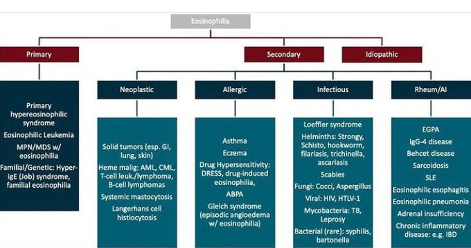
Let's explore the most important causes.
Eosinophilia - Hematology and Oncology
The Merck Manual provides a detailed, professional-level overview of eosinophilia, including its causes and evaluation.
Please read the following sections: Introduction (first few paragraphs): Note the definition (> 500/mcL) and the classification of severity (mild, moderate, severe). Etiology of Eosinophilia: This section highlights allergic disorders and parasitic infections as the most common causes. Table: Important Disorders and Treatments Associated With Eosinophilia: Skim this table to appreciate the breadth of causes. Pay particular attention to the 'Allergic or atopic disorders', 'Parasitic infections', and 'Systemic rheumatic diseases' categories. Evaluation of Eosinophilia: Focus on the 'History' section to see the key questions you must ask a patient with eosinophilia (travel, allergies, medications).
The "Big Two" and Other Key Causes
- Allergic/Atopic Disorders: This is the most common cause in developed countries. It includes conditions like allergic rhinitis, asthma, and atopic dermatitis (eczema). The eosinophilia is typically mild.
- Parasitic Infections: This is a major cause worldwide. The key is to think about tissue-invasive helminths (worms). Protozoa generally do not cause eosinophilia. This is why a travel history is absolutely critical.
- Drug Reactions: Many drugs can cause eosinophilia. A severe form is DRESS syndrome (Drug Reaction with Eosinophilia and Systemic Symptoms), a medical emergency characterized by fever, rash, eosinophilia, and internal organ involvement.
- Adrenal Insufficiency (Addison's Disease): A classic, albeit less common, cause. Glucocorticoids normally suppress eosinophil levels; their absence in adrenal insufficiency allows the count to rise.
- Malignancies: Especially Hodgkin lymphoma and certain myeloproliferative neoplasms.
- Autoimmune/Vasculitic Disorders: For example, Eosinophilic Granulomatosis with Polyangiitis (EGPA), formerly Churg-Strauss syndrome.
The following video provides some practical case examples.
TLC and DLC analysis | Dr Arvind Kumar
This clip from Dr. Arvind Kumar's lecture covers the main causes of eosinophilia and presents two illustrative cases.
Please watch from 10:00 to 12:24. Notice how the clinical presentation guides the diagnosis. The first case is classic allergic rhinitis. The second case, with its geographic clue ('patient from Bihar') and asthma-like symptoms, points towards tropical pulmonary eosinophilia, a parasitic reaction.
Test your understanding!
A 28-year-old medical student returns from a global health trip to Southeast Asia. A month later, she develops abdominal pain and diarrhea. Her CBC shows an AEC of 1,200/µL. What is the most likely category of illness, and what is a reasonable first diagnostic test?
Show answer
The combination of a recent travel history to a region where parasitic infections are endemic and the presence of moderate eosinophilia strongly suggests a helminthic (parasitic worm) infection. A reasonable first step in the workup would be to send stool samples for ova and parasite (O&P) testing.
Conclusion
Congratulations on completing our tour of the WBC differential! You are now equipped to tackle the most common abnormalities in neutrophils, lymphocytes, and eosinophils.
Key Takeaways:
- Always interpret absolute counts, not percentages.
- Lymphocytosis (>5,000/µL): Your differential is primarily reactive (viral) versus malignant (CLL). The patient's age, symptoms, and the absolute count itself guide your workup. Flow cytometry is the key test to distinguish between them.
- Eosinophilia (>500/µL): Your differential should be immediately anchored by allergies and parasites. A thorough history focusing on atopy, medications, and travel is the most important part of your evaluation.
Next Steps
We are now transitioning from the hematology lab to the chemistry lab. In our next lesson, we will begin our exploration of the Basic Metabolic Panel (BMP), a ubiquitous test you'll see every single day in the hospital. We will start by learning to recognize and interpret sodium abnormalities (hyponatremia and hypernatremia), a fundamental skill in managing fluid and electrolyte balance.