Hello! Welcome to the next lesson in our module on the liver and biliary system.
In our last session, we learned how to use gamma-glutamyl transferase (GGT) to confirm that an elevated alkaline phosphatase (ALP) is truly coming from the hepatobiliary system, allowing us to confidently identify a cholestatic pattern of injury. But what happens when that cholestatic process, or another liver issue, leads to jaundice? Your next diagnostic step is to figure out what kind of bilirubin is elevated.
Today, we will focus on how to interpret bilirubin fractionation to distinguish between conjugated and unconjugated hyperbilirubinemia. This is a critical skill that allows you to narrow down the causes of jaundice by localizing the problem: is it happening before the liver (pre-hepatic), within the liver (intra-hepatic), or after the liver (post-hepatic)?
Bilirubin Metabolism: The Foundation of Interpretation
To understand the different patterns of jaundice, we first need a clear picture of how bilirubin is processed in the body. It's a multi-step pathway, and a problem at any step leads to a distinct laboratory signature.
Let's start with a short video that provides a clear overview of bilirubin metabolism and defines the key terms we'll be using.
The Strong Medicine channel provides an excellent, concise review of the bilirubin pathway, from its creation to its excretion. This will set the foundation for the rest of our lesson.
Watch the first 2 minutes and 34 seconds of the video. Focus on the distinction between unconjugated (indirect) and conjugated (direct) bilirubin and their properties.
To summarize the key points from the video:
- Production: Bilirubin is a yellow pigment produced from the breakdown of heme, primarily from old red blood cells. This initial form is unconjugated bilirubin.
- Transport: Unconjugated bilirubin is lipid-soluble (not water-soluble) and must bind to albumin to travel through the blood to the liver.
- Conjugation: In the liver, the enzyme UGT (uridine diphosphoglucuronate glucuronosyltransferase) attaches glucuronic acid molecules to bilirubin. This converts it into conjugated bilirubin.
- Excretion: Conjugation makes bilirubin water-soluble. This allows it to be excreted from the liver into the bile, which then flows into the intestines.
In clinical practice, you will see these terms used interchangeably:
- Unconjugated Bilirubin ≈ Indirect Bilirubin
- Conjugated Bilirubin ≈ Direct Bilirubin
The key takeaway is that the liver's job is to convert water-insoluble, potentially toxic unconjugated bilirubin into a water-soluble form that can be safely excreted. The type of bilirubin that accumulates in the blood tells us where this process is failing.
Decoding the Patterns of Hyperbilirubinemia
When you see an elevated total bilirubin, the next step is to look at the "fractionation"—the breakdown between the direct (conjugated) and indirect (unconjugated) components. This will point you towards one of three main patterns. The Ninja Nerd video series offers a wonderfully detailed explanation of each.
1. Predominantly Unconjugated (Indirect) Hyperbilirubinemia
This pattern occurs when there is either overproduction of bilirubin, overwhelming the liver's capacity to conjugate it, or a defect in conjugation itself. This is often called a "pre-hepatic" problem.
Liver Function Tests (LFTs) | Clinical Medicine
This segment from the Ninja Nerd lecture on LFTs details the causes of unconjugated hyperbilirubinemia. It's a fantastic, in-depth look at the top differential diagnoses.
Watch the segment from 54:48 to 1:03:46. Pay close attention to the three main causes discussed (hemolysis, transport issues, and Gilbert's syndrome) and the key clinical finding regarding urine color.
As explained in the video, the main causes of unconjugated hyperbilirubinemia are:
- Hemolysis: Excessive breakdown of red blood cells (e.g., in sickle cell disease, autoimmune hemolytic anemia). Clues include anemia, a high LDH, and low haptoglobin.
- Impaired Hepatic Uptake/Conjugation:
- Gilbert's Syndrome: A very common, benign genetic condition causing reduced UGT enzyme activity. It presents as mild, transient jaundice during times of stress, illness, or fasting. The rest of the LFTs are normal.
- Crigler-Najjar Syndrome: A much rarer and more severe genetic absence of UGT activity.
- Drugs: Certain drugs, like rifampin, can impair the liver's uptake of bilirubin.
A crucial clinical pearl: because unconjugated bilirubin is not water-soluble, it cannot be filtered by the kidneys into the urine. Therefore, in isolated unconjugated hyperbilirubinemia, the urine color is normal. Stool color is also normal.
2. Predominantly Conjugated (Direct) Hyperbilirubinemia
This pattern indicates that the liver can conjugate bilirubin successfully, but it cannot excrete it properly. The conjugated, water-soluble bilirubin "backs up" and leaks into the bloodstream. This is a classic "post-hepatic" or obstructive pattern.
Liver Function Tests (LFTs) | Clinical Medicine
Let's continue with the Ninja Nerd video to understand the causes of conjugated hyperbilirubinemia.
Watch the segment from 1:09:07 to 1:18:51. Focus on how biliary obstruction (both intrahepatic and extrahepatic) leads to this pattern and the characteristic changes in urine and stool color.
The main causes of conjugated hyperbilirubinemia are:
- Biliary Obstruction: Something is physically blocking the bile ducts outside the liver (extrahepatic).
- Gallstones in the common bile duct (choledocholithiasis).
- Pancreatic cancer compressing the bile duct.
- Biliary strictures.
- Intrahepatic Cholestasis: Bile flow is impaired within the liver itself (e.g., Primary Biliary Cholangitis, cholestasis of pregnancy, drug-induced).
- Inherited Excretion Defects: Rare genetic disorders like Dubin-Johnson syndrome and Rotor syndrome.
The clinical pearls here are the opposite of the unconjugated pattern. Because conjugated bilirubin is water-soluble, it is excreted by the kidneys, causing dark, tea-colored urine. Furthermore, since bilirubin isn't reaching the gut, the stool lacks its normal brown pigment, leading to pale or clay-colored stools.
3. Mixed (Unconjugated and Conjugated) Hyperbilirubinemia
This pattern occurs when there is generalized liver dysfunction, or "intra-hepatic" disease. The damaged hepatocytes are impaired in their ability both to take up and conjugate bilirubin and to excrete it.
Liver Function Tests (LFTs) | Clinical Medicine
Finally, let's look at the pattern seen in direct liver cell damage.
Watch the short segment from 1:03:46 to 1:09:07. Note how hepatocellular injury (like hepatitis) causes elevations in both types of bilirubin, typically along with high AST and ALT.
This mixed pattern is the hallmark of hepatocellular injury, seen in conditions such as:
- Viral hepatitis (e.g., Hepatitis A, B, C)
- Alcoholic hepatitis
- Drug-induced liver injury
- Cirrhosis
This bilirubin pattern is almost always accompanied by the hepatocellular injury pattern we discussed previously: significantly elevated AST and ALT.
Consolidating the Differential Diagnosis
You have now seen the mechanisms and classic examples for each pattern. For a comprehensive written reference, the Medscape article on bilirubin provides excellent lists of causes.
Bilirubin: Reference Range, Interpretation, Collection and ...
This article from Medscape offers a detailed, text-based summary of the causes of hyperbilirubinemia. Reading this will reinforce what you learned in the videos and serve as a great future reference.
Read the sections titled 'Unconjugated hyperbilirubinemia' and 'Conjugated hyperbilirubinemia'. Skim the lists of causes to familiarize yourself with the breadth of the differential diagnosis for each pattern.
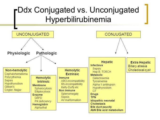
The flowchart below provides a great visual summary of how to categorize the causes of jaundice.
Test your understanding!
A 21-year-old medical student presents to student health services with mild yellowing of his eyes. He is studying for his final exams and has been pulling all-nighters and skipping meals. His lab results are:
- Total Bilirubin: 2.8 mg/dL (Normal: 0.3-1.0)
- Direct Bilirubin: 0.2 mg/dL (Normal: 0.1-0.3)
- ALT: 30 IU/L (Normal: 3-40)
- AST: 25 IU/L (Normal: 3-30)
- ALP: 80 IU/L (Normal: 30-100)
- Hemoglobin: 15 g/dL (Normal)
What is the pattern of hyperbilirubinemia, and what is the most likely diagnosis?
Show answer
The total bilirubin is elevated, but the direct (conjugated) bilirubin is normal. This means the excess is entirely indirect (unconjugated) bilirubin. The rest of his LFTs and his hemoglobin are normal, ruling out significant hepatocellular disease or hemolysis. This clinical picture of mild, isolated, unconjugated hyperbilirubinemia triggered by stress in a young person is classic for Gilbert's syndrome. This is a benign condition requiring no treatment, only reassurance.
A Practical Algorithm for Working Up Jaundice
Now, let's put it all together into a practical, step-by-step diagnostic algorithm. When a patient presents with jaundice, what do you do?
The Strong Medicine channel provides a fantastic, clinically-focused algorithm for working up a patient with jaundice based on their lab patterns.
Watch the segment from 8:55 to 12:05. This part directly translates the lab patterns we've discussed into concrete next diagnostic steps.
Here is a summary of that clinical workflow:
- See elevated total bilirubin.
- Fractionate it (check direct/indirect).
- Interpret the pattern and take the next step:
- Predominantly Unconjugated (Indirect): Think hemolysis or Gilbert's.
- Next Step: Check labs for hemolysis (CBC, reticulocyte count, LDH, haptoglobin). If negative, and the picture fits, consider Gilbert's.
- Predominantly Conjugated (Direct) with elevated ALP/GGT: Think biliary obstruction.
- Next Step: Order a right upper quadrant ultrasound to look for dilated bile ducts.
- Mixed, with elevated AST/ALT: Think hepatocellular disease.
- Next Step: Work up for causes of hepatitis (viral serologies, acetaminophen level, alcohol history, etc.) and consider an ultrasound.
- Predominantly Unconjugated (Indirect): Think hemolysis or Gilbert's.
Test your understanding!
A 68-year-old man presents with a week of jaundice, dark urine, and pale stools. His labs show:
- Total Bilirubin: 15.0 mg/dL
- Direct Bilirubin: 12.5 mg/dL
- ALT: 150 IU/L
- AST: 120 IU/L
- ALP: 600 IU/L
- GGT: 750 IU/L
What is the pattern, and what is the single most appropriate next diagnostic test?
Show answer
The pattern is a predominantly conjugated (direct) hyperbilirubinemia with a marked cholestatic picture (very high ALP and GGT). The clinical history of dark urine and pale stools strongly supports this. This entire picture points towards biliary obstruction. The most appropriate next test is a right upper quadrant ultrasound to look for evidence of bile duct dilation or a mass (like a pancreatic tumor) causing the obstruction.
Conclusion
By fractionating bilirubin, you can transform a general symptom like jaundice into a specific diagnostic pathway. Understanding these patterns is fundamental to interpreting liver function tests.
Key Takeaways:
- Unconjugated (Indirect) Hyperbilirubinemia: Caused by overproduction (hemolysis) or impaired conjugation (Gilbert's). Urine color is normal.
- Conjugated (Direct) Hyperbilirubinemia: Caused by impaired excretion or obstruction. Associated with dark urine, pale stools, and typically high ALP/GGT.
- Mixed Hyperbilirubinemia: Caused by hepatocellular damage (e.g., hepatitis, cirrhosis) and is typically seen with high AST/ALT.
- Your interpretation of the bilirubin pattern dictates your next diagnostic steps, whether it's checking for hemolysis or ordering an ultrasound.
So far, we have focused on markers of liver injury (AST/ALT) and cholestasis (ALP/GGT/Bilirubin). However, these tests don't tell us about the liver's actual functional capacity.
In our next lesson, we will complete our core LFT panel by learning how to assess hepatic synthetic function using albumin and the International Normalized Ratio (INR). These markers tell us how well the liver is performing its vital job of producing proteins.