Hello! Welcome back to our module on the liver and biliary system.
In our last lesson, we established the fundamental principle of differentiating liver injury into two main patterns: hepatocellular (marked by elevated ALT/AST) and cholestatic (marked by elevated ALP). We noted that while this is a powerful first step, alkaline phosphatase (ALP) isn't exclusive to the liver. This can create a common clinical dilemma: when you see a high ALP, how can you be certain it's coming from the biliary system and not from another source, like bone?
Today's lesson directly answers that question. We will focus on how to interpret gamma-glutamyl transferase (GGT) to investigate the origin of an elevated alkaline phosphatase. Mastering this will allow you to confidently distinguish between a liver problem and a bone problem, preventing unnecessary investigations and leading you to the correct diagnosis more efficiently.
The Specificity Problem with Alkaline Phosphatase (ALP)
As a quick review, ALP is an enzyme found on the surface of cells. In the liver, it's concentrated in the cells lining the bile ducts (biliary epithelium). When bile flow is blocked (cholestasis), the synthesis of ALP is induced, and levels in the blood rise.
However, the liver is not the only source. To understand why we need another test, let's look at the other major contributors to serum ALP.
Liver Function Test Interpretation (LFTs) | Liver Enzymes Remastered (Bilirubin, GGT, Alk Phos)
The MedCram video provides a concise explanation of why ALP has low specificity for liver disease and lists the other common causes of its elevation.
Watch the segment from 3:48 to 5:42. Pay close attention to the three main categories that can cause an elevated alkaline phosphatase.
As the video highlights, a high ALP can be due to:
- Cholestasis: The hepatobiliary cause we are interested in.
- Bone Disease: Specifically, conditions with high osteoblastic activity (bone formation), such as Paget's disease, healing fractures, or bone metastases (especially blastic ones like from prostate cancer). Bone growth in children and adolescents is also a normal cause of elevated ALP.
- Pregnancy: The placenta produces its own ALP, leading to elevated levels, particularly in the third trimester.
Given these other significant sources, an isolated elevation in ALP requires a tie-breaker test to confirm its origin. This is where GGT comes in.
GGT: The Confirmatory Test
Gamma-glutamyl transferase (GGT) is another enzyme found in high concentrations in the liver and biliary system. Like ALP, its levels rise in cholestasis. However, crucially, GGT is not significantly elevated in bone disease.
This difference in tissue distribution is the key to its clinical utility. By measuring GGT alongside a high ALP, you can determine the source of the elevation.
The interpretation is straightforward:
- Elevated ALP + Elevated GGT: The problem is almost certainly in the hepatobiliary system.
- Elevated ALP + Normal GGT: The problem is not in the hepatobiliary system. You should investigate extra-hepatic causes, most commonly bone disease.
Let's read a clear, written summary of this essential relationship.
Interpretation of Liver Function Tests (LFTs)
The Geeky Medics guide provides an excellent, concise summary of how to interpret ALP and GGT together.
Read the section titled 'ALP / GGT'. Focus on the bullet points that describe how the two tests are interpreted together to localize the source of a raised ALP.
This principle is one of the most reliable rules in clinical chemistry. The American Association for the Study of Liver Diseases (AASLD) formalizes this in their guidelines, stating that an "isolated ALP elevation warrants GGT testing to confirm the source" (from the article "How to approach elevated liver enzymes?").
The Clinical Algorithm for an Elevated ALP
Now, let's visualize how this works in practice. When you're faced with an elevated ALP on a lab report, you should follow a systematic approach. The flowchart below illustrates this diagnostic pathway perfectly.
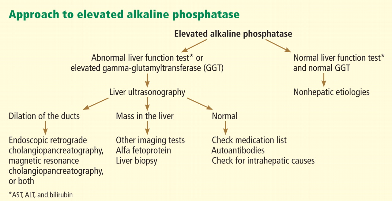
Let's walk through the logic of this chart:
- First, you see an elevated ALP.
- Next, you check the GGT.
- If GGT is elevated (along with the ALP), you are confident the cause is hepatobiliary. Your next step, as the chart shows, is typically a liver ultrasound to look for signs of biliary duct dilation (suggesting an obstruction) or other liver parenchymal abnormalities.
- If GGT is normal, you can confidently rule out a significant cholestatic liver process. You should then consider non-hepatic causes, primarily bone disease.
Test your understanding!
A 72-year-old woman is evaluated for persistent back pain. Her lab results are:
- ALT: 25 IU/L (Normal: 3-40)
- AST: 28 IU/L (Normal: 3-30)
- ALP: 350 IU/L (Normal: 30-100)
- Bilirubin: 12 µmol/L (Normal: 3-17)
You add on a GGT, which comes back at 45 IU/L (Normal: 8-60).
What is the most likely source of her elevated ALP, and what general area would you investigate next?
Show answer
The ALP is significantly elevated, but the GGT is normal, as are the other LFTs. This pattern strongly indicates a non-hepatobiliary source for the elevated ALP. Given her age and complaint of back pain, a bony process (such as Paget's disease, healing fracture, or metastatic disease) is the most likely cause. The next steps should focus on investigating her bone pathology, not her liver.
The Special Case: Isolated GGT Elevation
What if you see the reverse pattern: a normal ALP but a high GGT?
An isolated GGT elevation is a sensitive, but not very specific, marker. However, it is most classically associated with two things:
- Alcohol Consumption: GGT is induced by alcohol, so it will be elevated in individuals who drink regularly, even before significant liver damage occurs.
- Enzyme-Inducing Drugs: Certain medications (e.g., some anticonvulsants like phenytoin and carbamazepine) can also cause an isolated rise in GGT.
For a final review, let's watch a video that ties these concepts together with clinical cases.
Liver and pancreatic enzymes explained | AST, ALT, GGT, ALP, Amylase & Lipase
This Medicosis Perfectionalis video concisely explains the relationship between ALP and GGT and includes case examples that highlight the patterns we've discussed.
Watch the two short clips: From 1:54 to 2:48, which directly explains the ALP/GGT relationship for differentiating biliary vs. bone sources. From 8:30 to 9:00, which presents a case of alcoholic fatty liver, a classic cause of elevated GGT.
Conclusion
You have now added a crucial tool to your LFT interpretation toolkit. By using GGT as a confirmatory test, you can solve the ambiguity of an elevated ALP and confidently determine whether the pathology lies within the hepatobiliary system or elsewhere.
Key Takeaways:
- Alkaline Phosphatase (ALP) is elevated in both cholestasis and various bone diseases.
- Gamma-glutamyl transferase (GGT) is elevated in cholestasis but not in bone disease.
- Elevated ALP + Elevated GGT points to a hepatobiliary cause. The next step is usually imaging (e.g., ultrasound).
- Elevated ALP + Normal GGT points to a non-hepatobiliary cause, most commonly from bone.
- An isolated elevation in GGT is often associated with alcohol use or certain medications.
Now that we can confidently identify a cholestatic pattern, our next step is to further characterize it. In cholestasis, bilirubin can also be elevated.
In our next lesson, we will dive into bilirubin fractionation. You will learn to distinguish between conjugated (direct) and unconjugated (indirect) hyperbilirubinemia, which will allow you to further localize the problem—whether it's before the liver (pre-hepatic), within the liver (hepatic), or after the liver (post-hepatic).