Hello! Welcome back to our module on "Inflammatory and Cardiac Markers."
In our previous lesson, we explored the interpretation of general inflammatory markers, CRP and ESR. You learned to distinguish between these "fast" and "slow" reactants to assess for acute and chronic inflammation. Today, we transition from general systemic markers to a highly specific and critical biomarker used in cardiology: cardiac troponin.
Your learning outcome for this lesson is to interpret troponin levels in the diagnosis and risk stratification of acute coronary syndrome (ACS). We will cover what troponin is, how its levels change during a heart attack, and, most importantly, how to use this powerful test to make accurate diagnoses and guide urgent clinical decisions. This is one of the most high-stakes lab tests you will encounter, and mastering its interpretation is essential.
1. What is Cardiac Troponin?
Troponin is a protein complex essential for muscle contraction. While it exists in all muscle tissue, certain isoforms—cardiac Troponin I (cTnI) and cardiac Troponin T (cTnT)—are unique to the heart muscle.
When heart muscle cells (myocytes) are damaged and die (a process called necrosis), their contents leak into the bloodstream. Because cTnI and cTnT are specific to the heart, their presence in the blood is a direct indicator of myocardial injury. This makes troponin the cornerstone for diagnosing a myocardial infarction (MI), or heart attack.
The progression of an MI involves a characteristic rise and fall of troponin levels.
Acute Coronary Syndromes - SAEM
This table from a Society for Academic Emergency Medicine (SAEM) resource shows the typical time course of cardiac biomarkers after an MI. Notice how long troponins can remain elevated compared to older markers like CK-MB.
Review 'Table 1. Time Course of Cardiac Biomarkers' in the section on 'Enzyme testing'. Focus on the initial elevation, peak, and return to baseline for Troponin I and Troponin T. This long duration of elevation is why a patient can still be diagnosed with an MI days after their symptoms began.
The Advent of High-Sensitivity Troponins (hs-cTn)
In recent years, high-sensitivity cardiac troponin (hs-cTn) assays have become the standard. It's important to understand what "high-sensitivity" means in this context. It does not refer to a new type of troponin, but rather a more advanced test.
- Greater Analytical Sensitivity: These assays can detect much lower concentrations of troponin in the blood.
- Greater Precision: They provide more reliable and reproducible results at these very low levels.
This advancement has revolutionized how we evaluate patients with chest pain.
Learn to Love High Sensitivity Troponins | The EM & Acute Care Course
To clarify the difference between conventional and high-sensitivity troponins, watch this brief explanation.
Watch the first 1 minute and 28 seconds. The key concept is that hs-cTn assays simply have a much lower threshold of detection, allowing us to identify myocardial injury that was previously missed.
2. Diagnosing Acute Coronary Syndrome (ACS)
Acute Coronary Syndrome is a spectrum of conditions caused by a sudden reduction in blood flow to the heart. It includes:
- ST-Elevation Myocardial Infarction (STEMI): A complete blockage of a coronary artery, causing characteristic ST-segment elevation on the ECG.
- Non-ST-Elevation Myocardial Infarction (NSTEMI): A partial or intermittent blockage causing myocardial necrosis, but without ST-elevation on the ECG.
- Unstable Angina (UA): A partial blockage causing ischemic symptoms, but without enough damage to cause significant myocardial necrosis and a rise in troponin.
Troponin is the key lab test that differentiates NSTEMI from Unstable Angina.
Your Easy Guide to Acute Coronary Syndrome
Let's watch a concise overview that defines the components of ACS and highlights troponin's central diagnostic role.
Watch the first 1 minute and 7 seconds. Focus on the single factor that separates NSTEMI from unstable angina: the presence of a troponin rise.
Myocardial Injury vs. Myocardial Infarction: The "Delta"
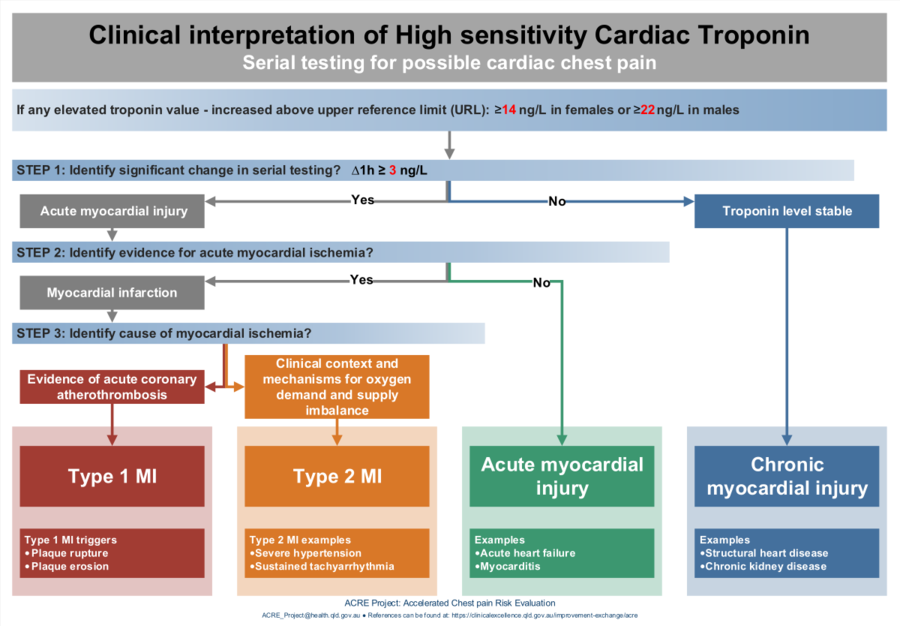
With hs-cTn assays, we can detect very small amounts of troponin, even in healthy individuals. The cutoff for abnormality is the 99th percentile upper reference limit (URL)—the highest level seen in 99% of a healthy reference population.
- Myocardial Injury: Is defined as any troponin value above the 99th percentile URL.
- Acute Myocardial Infarction (MI): Requires evidence of acute myocardial ischemia in addition to the injury. This is established by observing a dynamic rise and/or fall in troponin values on serial testing, known as the "delta," in a patient with symptoms or ECG changes suggesting ischemia.
This "delta" is critical. A single elevated troponin tells you there is heart muscle injury, but it doesn't tell you if it's new or old. Seeing the level change over a short period (e.g., 1-3 hours) confirms the process is acute.
High-Sensitivity Cardiac Troponin and the 2021 AHA/ACC/ ...
Let's explore this crucial concept of 'delta troponin' more formally. This expert document explains the principles behind using serial measurements.
Read the section titled 'Information About a Changing Pattern of Values (Deltas)'. Pay close attention to Figure 4, which visually contrasts the troponin patterns in acute versus chronic myocardial injury.
3. Risk Stratification in NSTEMI and Unstable Angina
Once a diagnosis of NSTEMI or UA is suspected, troponin levels—along with clinical features—are used to risk-stratify the patient. This helps determine the urgency and type of intervention needed. Clinical risk scores like TIMI and GRACE incorporate troponin values to predict the risk of major adverse cardiac events.
A higher troponin peak indicates a larger area of infarction and is associated with a worse prognosis. This information helps clinicians decide whether a patient requires an urgent invasive strategy (i.e., cardiac catheterization) or can be managed more conservatively.
Your Easy Guide to Acute Coronary Syndrome
This video segment explains how risk scores are used in NSTEMI and UA to guide management decisions, specifically the timing of cardiac catheterization.
Watch from 04:58 to 07:32. You don't need to memorize the scoring systems, but understand the principle: higher-risk patients (often with higher troponins or dynamic ECG changes) benefit from an earlier invasive approach.
4. The Broad Differential of an Elevated Troponin
This is one of the most important practical challenges you will face. With the widespread use of hs-cTn assays, you will frequently encounter elevated troponins in patients who are not having a classic heart attack. It is crucial to distinguish a Type 1 MI from other causes of myocardial injury.
- Type 1 MI: The classic ACS event caused by atherosclerotic plaque rupture and coronary thrombosis. This is what requires urgent cardiology consultation, antiplatelet therapy, anticoagulation, and potential catheterization.
- Type 2 MI: Myocardial injury and infarction due to a supply-demand mismatch. The coronary arteries themselves are not acutely blocked. Examples include:
- Decreased Supply: Severe anemia, respiratory failure, hypotension/shock.
- Increased Demand: Sepsis, tachyarrhythmias (like rapid atrial fibrillation), severe hypertension.
- The treatment for Type 2 MI is to correct the underlying problem, not to give ACS medications.
- Chronic Myocardial Injury: A stable, non-dynamic elevation in troponin seen in conditions like chronic kidney disease (CKD), stable heart failure, or pulmonary hypertension. This is due to chronic cardiac stress, not an acute event.
Your Easy Guide to Acute Coronary Syndrome
Differentiating these conditions is fundamental to safe and effective practice. This video provides an excellent explanation of how to approach this differential diagnosis.
Watch from 15:41 to 19:35. The most important takeaway is that Type 2 MI and chronic myocardial injury are NOT treated with the standard ACS protocol. Misdiagnosing a Type 2 MI as a Type 1 MI can lead to harmful and unnecessary interventions.
Even when a troponin elevation is determined to be chronic (e.g., in a patient with end-stage renal disease), it is not benign. It signifies underlying cardiac stress and carries a poor long-term prognosis.
Test your understanding!
A 74-year-old man with a history of hypertension and diabetes is admitted to the ICU with pneumonia and septic shock. His blood pressure is low, and he is tachycardic. A routine lab draw shows an initial troponin of 100 ng/L (99th percentile URL is 14 ng/L). A repeat level 3 hours later is 500 ng/L. His ECG shows tachycardia but no ST elevations.
Based on these findings, what is the most likely diagnosis, and what is the primary management strategy?
Show answer
The most likely diagnosis is a Type 2 Myocardial Infarction.
Reasoning: The patient has a clear reason for a supply-demand mismatch (septic shock causing hypotension and tachycardia). While he has a dynamic rise in troponin, indicating acute myocardial injury, it is in the context of a severe systemic illness. The primary management is to treat the underlying condition: aggressively manage the septic shock with fluids, vasopressors, and antibiotics. He should not be started on dual antiplatelet therapy or sent for an urgent cardiac catheterization based on this information alone. The goal is to restore hemodynamic stability to reduce the strain on his heart.
Conclusion
You have now learned to interpret one of the most vital tests in acute care medicine. Understanding the nuances of troponin will allow you to confidently diagnose myocardial infarction, risk-stratify patients, and, just as importantly, avoid misattributing all troponin elevations to acute coronary syndrome.
Key Takeaways:
- Troponin is the gold standard for diagnosing myocardial infarction due to its cardiac specificity.
- A dynamic rise and/or fall (delta) is the key to differentiating an acute MI from chronic myocardial injury.
- High-sensitivity troponins allow for earlier and more frequent detection of injury, which enables rapid rule-out protocols but also increases the detection of non-ACS related elevations.
- Always consider the clinical context. The most critical step in interpreting an elevated troponin is to differentiate a Type 1 MI (plaque rupture) from a Type 2 MI (supply-demand mismatch) and chronic injury, as their management is fundamentally different.
In our next lesson, we will continue our focus on cardiac biomarkers by examining B-type natriuretic peptide (BNP) and its role in the diagnosis and management of heart failure.