Hello! Welcome to a new module focused on "Inflammatory and Cardiac Markers."
In our last session, we concluded the module on Coagulation Studies by learning how to perform a comprehensive, evidence-based assessment of a patient's bleeding risk before an invasive procedure. You learned that this is a clinical process driven by history and physical exam, with laboratory tests used selectively, not as a routine screen.
Today, we begin our new module by looking at two of the most frequently ordered tests in medicine used to detect inflammation: the C-reactive protein (CRP) and the erythrocyte sedimentation rate (ESR). Your learning outcome for this lesson is to interpret C-reactive protein (CRP) and erythrocyte sedimentation rate (ESR) in inflammatory conditions. We will explore what these markers represent, how they differ, and most importantly, develop a practical framework for when to use each test—or both—to guide your clinical judgment.
1. The Acute Phase Response: Your Body's Systemic Alarm
When the body encounters an inflammatory stimulus—be it an infection, trauma, or an autoimmune flare-up—it mounts a systemic reaction called the acute phase response. A key part of this response is the liver, which, triggered by inflammatory cytokines like Interleukin-6 (IL-6), dramatically changes its protein production. It ramps up the synthesis of some proteins, called positive acute phase reactants, and decreases others, known as negative acute phase reactants.
This fundamental concept is the basis for understanding both CRP and ESR.
Acute Phase Reactants (APRs) - ESR and CRP - Inflammation markers - Lab test results
To get a feel for this process, let's watch a segment from Medicosis Perfectionalis. This will introduce the concept of acute phase reactants and explain why the liver shifts its production priorities during inflammation.
Watch from 03:08 to 09:45. Focus on the distinction between positive and negative acute phase reactants and the rationale behind this shift in protein synthesis.
As you saw, the body increases proteins that help the immune system (like CRP and fibrinogen) while conserving resources by decreasing others (like albumin and transferrin). This leads us to our two key markers:
- C-Reactive Protein (CRP): A positive acute phase reactant itself. Measuring CRP is a direct measurement of this inflammatory response.
- Erythrocyte Sedimentation Rate (ESR): A physical measurement that is indirectly influenced by the concentration of acute phase reactants, especially fibrinogen.
2. C-Reactive Protein (CRP): The Fast Responder
CRP is a protein that directly participates in the innate immune response by binding to pathogens and damaged cells, marking them for destruction. From a clinical perspective, its most important features are its kinetics.
Pathophysiology and Kinetics:
- Direct Marker: Produced by the liver in direct response to IL-6.
- Rapid Rise: Levels begin to increase within 4-6 hours of an inflammatory stimulus.
- Rapid Fall: Has a short half-life of about 19 hours. This means that if the inflammation is treated effectively, CRP levels will normalize quickly (within days).
This "fast on, fast off" characteristic makes CRP an excellent marker for detecting acute inflammation and for monitoring the response to therapy (e.g., seeing if antibiotics are working for a bacterial infection).
Making sense of inflammatory markers
The document 'Making sense of inflammatory markers' provides a great overview of how to interpret CRP levels.
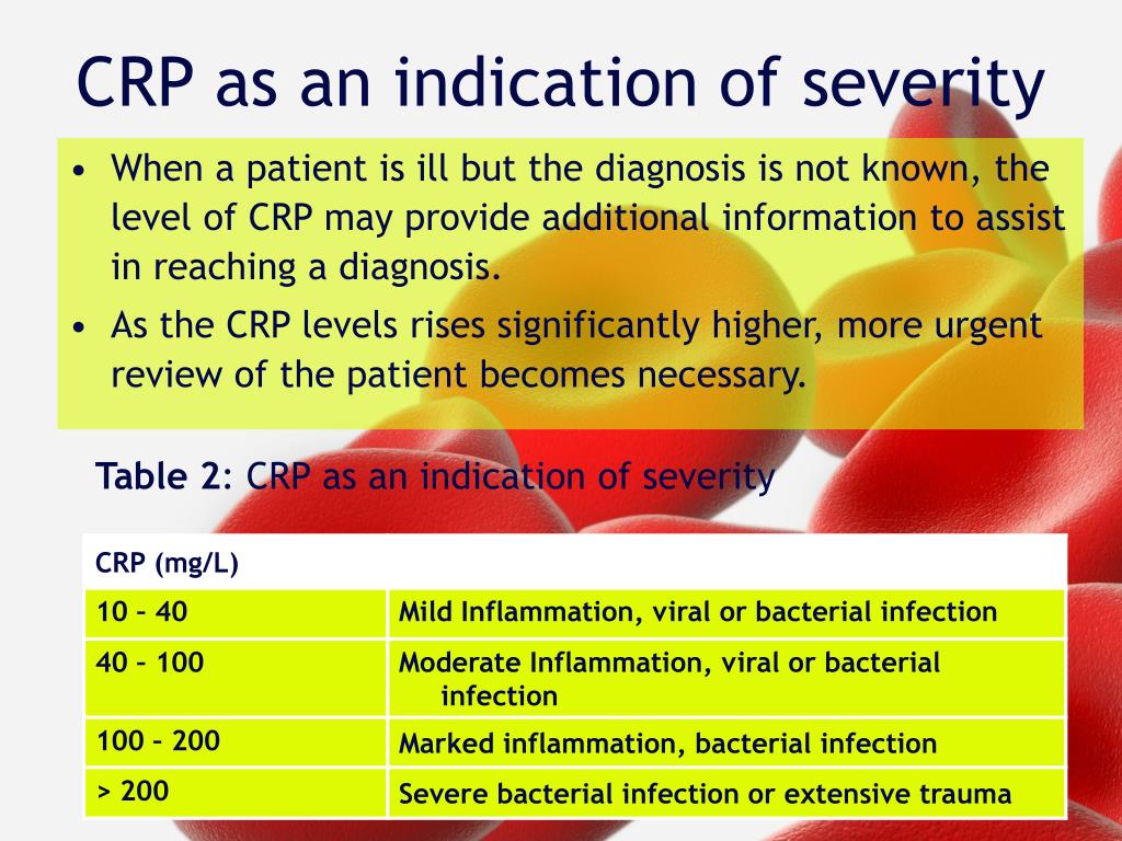
Read the section titled 'Measurement and interpretation of CRP levels'. Pay close attention to the general rules for interpreting the magnitude of the CRP rise and the important exceptions where CRP might be misleadingly low (like in SLE or ulcerative colitis).
The magnitude of the CRP elevation can give you a clue about the severity or type of inflammation.
3. Erythrocyte Sedimentation Rate (ESR): The Slow and Steady Marker
Unlike CRP, the ESR is not a measure of a specific molecule. It's a physical test that measures how quickly red blood cells (erythrocytes) settle in a vertical tube of blood over one hour.
Pathophysiology and Kinetics:
- Indirect Marker: In an inflammatory state, high levels of acute phase proteins (especially fibrinogen) neutralize the negative surface charge of red blood cells. This allows them to stick together in stacks called 'rouleaux'. These stacks are denser and fall faster, resulting in a higher ESR.
- Slow Rise: The ESR rises slowly, typically over 24-48 hours, because it depends on the liver producing proteins like fibrinogen.
- Slow Fall: Because fibrinogen has a long half-life (several days), the ESR can remain elevated for days or even weeks after the inflammation has resolved.
What you need to know about your ESR lab result
This video from Connected Rheumatology provides a clear explanation of the ESR test and what influences the result.
Watch from 00:44 to 04:11. This will help you visualize the process of rouleaux formation and understand the many non-inflammatory factors that can affect the ESR, such as age, gender, and anemia.
The ESR's slow kinetics make it less useful for tracking acute changes. However, its stability makes it valuable for diagnosing and monitoring certain chronic inflammatory diseases, such as:
- Polymyalgia Rheumatica (PMR)
- Giant Cell Arteritis (GCA)
- Rheumatoid Arthritis (RA)
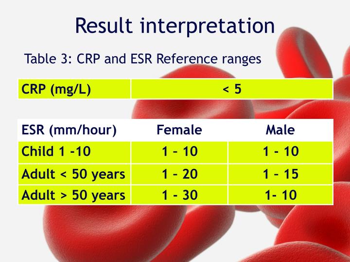
A key clinical pearl is that a markedly elevated ESR (e.g., > 100 mm/hr) is almost always pathological and should prompt a thorough investigation for infection, malignancy, or a significant rheumatological condition.
4. Head-to-Head: Choosing the Right Test
Now for the most practical question: which test should you order and when? The answer depends entirely on the clinical question you are asking.
The key difference lies in their half-lives.
Acute Phase Reactants (APRs) - ESR and CRP - Inflammation markers - Lab test results
Let's return to the Medicosis Perfectionalis video for a direct and memorable comparison of ESR and CRP, focusing on their differing kinetics.
Watch from 09:45 to 12:47. The mnemonic here is helpful: C-Reactive Protein is fast and flexible, while Sedimentation Rate is slow and less dynamic.
Here's a summary of the recommendations, based on guidelines from the College of American Pathologists.
C-Reactive Protein and Erythrocyte Sedimentation Rate Test Use
This document from the College of American Pathologists provides clear, evidence-based recommendations on the use of ESR and CRP.
Read the sections 'ESR and CRP Testing Recommendations' and 'INSIGHTS'. This will give you the formal guidelines on when CRP alone is preferred, when ESR remains useful, and when ordering both is beneficial.
| Characteristic | C-Reactive Protein (CRP) | Erythrocyte Sedimentation Rate (ESR) |
|---|---|---|
| What it measures | A specific acute phase protein | Rate of RBC settling (indirectly reflects plasma proteins) |
| Kinetics | Fast: Rises in 4-6 hrs, half-life 19 hrs | Slow: Rises in 24-48 hrs, half-life days-weeks |
| Primary Use | Detecting acute inflammation; Monitoring acute treatment response | Diagnosing/monitoring specific chronic inflammatory diseases |
| Confounders | Obesity | Age, gender, anemia, pregnancy, renal failure, globulins |
| Recommendation | Preferred test for initial evaluation of acute inflammation. | Useful for GCA, PMR, RA. Not a good screening test. |
When should you order both?
While routine ordering of both is discouraged, there are specific clinical situations where their discordant patterns provide valuable diagnostic information:
- Systemic Lupus Erythematosus (SLE): In an SLE flare, ESR is often very high while CRP is low or normal. If both are high, you should have a higher suspicion for a superimposed infection.
- Giant Cell Arteritis (GCA): Using both tests increases diagnostic sensitivity, as some patients may have a normal ESR but an elevated CRP.
- Orthopedic Infections: Especially in prosthetic joint infections, the combination of tests can improve diagnostic accuracy.
Test your understanding!
A 45-year-old woman with a 5-year history of rheumatoid arthritis (RA) comes to your clinic. She feels her joints are more swollen and painful over the last month. You want to assess her disease activity.
A separate patient, a 65-year-old man, is admitted to the hospital with high fever, cough, and shortness of breath. You suspect bacterial pneumonia and start him on IV antibiotics. You want to monitor his response to treatment over the next 48-72 hours.
For which patient is the ESR a more appropriate marker to track their condition, and for which is the CRP better? Why?
Show answer
- For the woman with rheumatoid arthritis, the ESR (along with CRP) is a very useful marker. RA is a chronic inflammatory condition, and the ESR's slow, stable nature is well-suited to monitoring long-term disease activity.
- For the man with suspected bacterial pneumonia, the CRP is the superior marker for monitoring short-term treatment response. Because of its short half-life, a significant drop in the CRP level within 48-72 hours would provide strong evidence that the antibiotics are effective. The ESR would still be elevated due to its slow kinetics and would not be helpful for this acute question.
Conclusion
You have now learned to differentiate between CRP and ESR, two fundamental markers of inflammation. By understanding their distinct physiological roles and kinetic properties, you can select the right test for the right clinical question, avoiding unnecessary testing and interpreting the results with more confidence.
Key Takeaways:
- CRP is the "fast" marker: It rises and falls quickly, making it ideal for detecting acute inflammation and monitoring the response to acute therapies.
- ESR is the "slow" marker: It rises and falls slowly, making it useful for diagnosing and monitoring specific chronic inflammatory conditions like GCA, PMR, and RA.
- Always consider the context: Both are non-specific tests. Their results must be interpreted alongside the patient's history, physical exam, and other laboratory findings.
- Order wisely: For a general screen for acute inflammation, CRP alone is usually sufficient. Reserve ordering both for specific clinical scenarios (like suspected GCA or differentiating an SLE flare from infection) where their differing patterns can provide diagnostic clues.
In our next lesson, we will shift our focus from these general markers of inflammation to a highly specific and critical biomarker used in cardiology. We will learn how to interpret troponin levels in the diagnosis and risk stratification of acute coronary syndrome.