Welcome back! In our previous lessons, we explored the final step of a hormone's journey: how it delivers its message to a target cell. We distinguished between lipid-soluble hormones like steroids, which enter the cell to act on DNA directly, and water-soluble peptide hormones, which use second messenger systems like cAMP and IP3 to signal from the cell surface.
Now, we will rewind to the beginning of that journey. This lesson addresses the fundamental questions of where hormones come from and how they travel. We will cover the general principles of hormone synthesis, secretion, and transport in the blood. You will learn how the chemical nature of a hormone—whether it's a protein or a steroid—dictates its entire lifecycle, from creation to its journey through the bloodstream. This is a critical foundation for understanding hormonal imbalances in conditions like PCOS.
1. Hormone Synthesis: The "Factories"
Hormone synthesis is the process of creating hormone molecules within an endocrine cell. The method of synthesis depends entirely on the hormone's chemical class. We will focus on the two most important classes for our course: peptide/protein hormones and steroid hormones.
A. Synthesis of Peptide and Protein Hormones
Peptide hormones, such as insulin, LH, and FSH, are essentially small proteins. Their synthesis follows the central dogma of molecular biology: the genetic code in DNA is used to build a protein. Since your background may not have focused heavily on this, let's break it down step-by-step. The process is like an assembly line within the cell.
- Transcription: A specific gene on the DNA in the cell's nucleus is read and copied into a messenger RNA (mRNA) molecule.
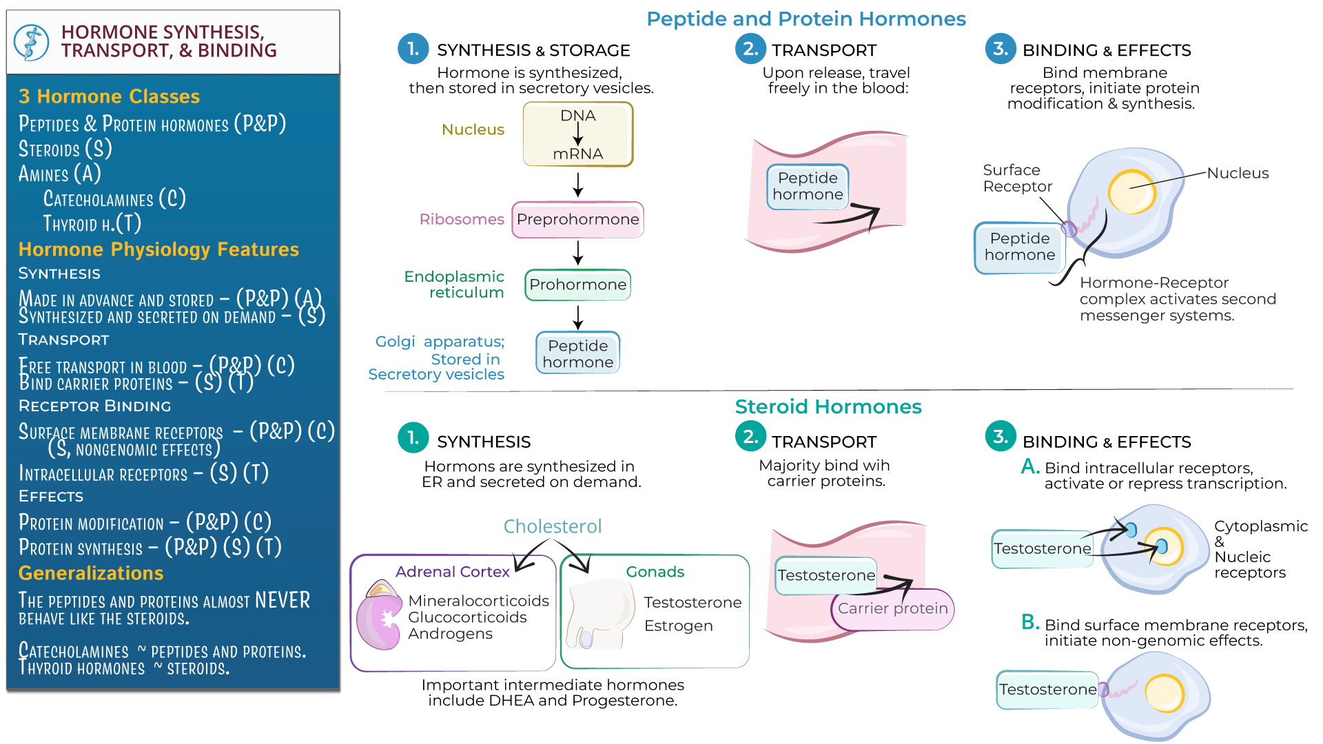
- Translation: The mRNA molecule travels out of the nucleus to the rough endoplasmic reticulum (ER), where ribosomes read the mRNA code and assemble a chain of amino acids. This initial, large, inactive version is called a preprohormone.
- Processing in the ER: While still in the ER, a "signal peptide" sequence is cut off, converting the molecule into a smaller, but still inactive, prohormone.
- Packaging in the Golgi Apparatus: The prohormone is transported to the Golgi apparatus, where it is packaged into small, membrane-bound sacs called secretory vesicles. Inside these vesicles, enzymes chop the prohormone into the final, biologically active hormone and other peptide fragments.
- Storage: These vesicles, filled with active hormone, are stored within the cell, ready for release.
The following image provides a clear visual summary of this intricate process.

To solidify your understanding of this pathway, please read the detailed explanation provided in the following resource.
Principles of endocrinology - NCBI - NIH
The textbook 'Endocrinology: An Integrated Approach' offers a clear, step-by-step description of protein and peptide hormone synthesis. This will reinforce the concepts we've just outlined.
Please read the section titled 'Hormone synthesis'. Focus on the detailed description of the synthesis of protein and peptide hormones, from the transcription of a gene to the final packaging into secretory granules. You can skim the parts on steroid and amine hormones for now, as we'll cover those next.
B. Synthesis of Steroid Hormones
Steroid hormones—including estrogens, progesterone, and androgens like testosterone—are not built from a genetic blueprint like proteins. Instead, they are chemically modified from a common precursor molecule: cholesterol.
The process is enzymatic, not genetic. A series of specialized enzymes within the cell's mitochondria and smooth endoplasmic reticulum modify the cholesterol molecule in a step-by-step fashion.
- Precursor: All steroid hormones originate from the 27-carbon lipid, cholesterol.
- Rate-Limiting Step: The first and most crucial conversion is from cholesterol to a molecule called pregnenolone. This step is the main point of regulation.
- Specialization: The specific steroid hormone produced by a cell (e.g., cortisol in the adrenal gland vs. testosterone in the ovary) depends on which specific set of enzymes that cell possesses.
This video provides an excellent introduction to this concept.
The video 'Synthesis of steroid hormones' by Andrey K clearly explains that all steroid hormones are derived from cholesterol and introduces the enzymatic pathways involved.
Watch the segment from 03:14 to 05:07. Focus on understanding that cholesterol is the universal precursor and that the first conversion to pregnenolone is the key rate-limiting step. Don't worry about memorizing the names of all the subsequent enzymes or pathways shown; the principle is what matters.
2. Hormone Secretion: Release into the Bloodstream
Once synthesized, hormones must be released from the cell. Again, the mechanism depends on the hormone's type.
-
Peptide Hormones: Because they are stored in vesicles, their release is a highly regulated process called exocytosis. When the cell receives a stimulus (e.g., GnRH stimulating the pituitary), the vesicles move to the cell membrane, fuse with it, and release their contents into the bloodstream. They are secreted in "bursts" or "pulses" when needed.
-
Steroid Hormones: Steroids are lipid-soluble. This means they cannot be stored in membrane-bound vesicles because they would simply diffuse right through the lipid membrane. Therefore, they are synthesized on demand. When a cell is stimulated to produce a steroid, it starts the enzymatic process, and the newly made steroid hormones immediately diffuse across the cell membrane into the blood. Their secretion rate is therefore controlled by regulating their synthesis rate.
3. Hormone Transport: The Journey in the Blood
After secretion, hormones travel through the circulatory system to reach their target tissues. Their solubility in blood (which is mostly water) determines how they travel.
A. Water-Soluble vs. Lipid-Soluble Hormones
-
Water-Soluble (Hydrophilic): Peptide hormones and catecholamines (like epinephrine) dissolve easily in the blood plasma and circulate in a free, unbound state.
-
Lipid-Soluble (Hydrophobic): Steroid hormones and thyroid hormones do not dissolve well in water. To travel in the bloodstream, they must be attached to water-soluble transport proteins (or carrier proteins). These proteins, mostly produced by the liver, act like chaperones, carrying the hormone through the blood.
Watch this brief clip that introduces the need for carrier proteins.
Let's revisit the 'Synthesis of steroid hormones' video. The beginning of this video provides a perfect explanation of why steroid hormones need carrier proteins to travel in the blood.
Please watch from 00:53 to 01:46. Pay attention to the problem of hydrophobicity and the solution: binding to carrier proteins like albumin and specific globulins.
B. Bound vs. Free Hormone
For hormones that use transport proteins, an equilibrium exists in the blood between the hormone that is attached to a protein (bound) and the small fraction that is not (free).
- Free Hormone: Only the free, unbound hormone is considered biologically active. This is because only the free hormone can leave the bloodstream, diffuse into a target tissue, and bind to its receptor.
- Bound Hormone: The bound hormone acts as a circulating reservoir. As free hormone is used up by tissues, some bound hormone is released from its carrier protein to replenish the free pool.
This binding also protects the hormone from being broken down by enzymes in the liver and filtered by the kidneys. As a result, protein-bound hormones have a much longer half-life (the time it takes for half of the hormone to be removed from the blood) than free-circulating hormones. For example, the half-life of cortisol (highly bound) is hours, while that of epinephrine (free) is seconds.
The concept of binding proteins is extremely important in PCOS. For example, Sex Hormone-Binding Globulin (SHBG) is the primary transport protein for testosterone. Conditions that lower SHBG levels, like the high insulin levels often seen in PCOS, lead to a higher proportion of free, active testosterone, contributing to symptoms of hyperandrogenism even if total testosterone levels are not dramatically elevated.
The following reading provides an excellent summary of hormone transport, binding proteins, and the significance of the free hormone fraction.
Principles of endocrinology - NCBI - NIH
The 'Principles of endocrinology' text provides a concise yet thorough explanation of hormone transport in the circulation, which ties together all the concepts we have just discussed.
Please read the section titled 'Transport of hormones in the circulation and their half-lives'. Focus on the role of binding proteins, the equilibrium between bound and free hormones, and how this affects biological activity and half-life.
Test your understanding!
A patient has a liver condition that significantly reduces its ability to produce transport proteins, including SHBG. How would this likely affect the patient's levels of total testosterone versus free testosterone? Which of these changes is more likely to cause clinical symptoms?
Show answer
With fewer SHBG molecules available, less testosterone can be bound in the bloodstream. This means a larger proportion of the total testosterone will be in the free, active form. The body might compensate by reducing overall testosterone production, so the total testosterone level could decrease or stay the same. However, the free testosterone level would likely increase. The increase in free testosterone is what would cause clinical symptoms (like hirsutism or acne), as this is the biologically active form that acts on tissues.
Summary Chart
This image provides an excellent side-by-side comparison of the entire lifecycle of peptide versus steroid hormones, summarizing this lesson and linking it back to the previous ones on hormone action.
Conclusion
In this lesson, we have traced the life of a hormone from its synthesis to its transport through the body. We've established a crucial dichotomy based on chemical structure that governs this entire process.
Key Takeaways:
- Peptide/Protein Hormones (e.g., LH, FSH, insulin) are built from amino acids via gene transcription and translation. They are stored in vesicles, released on demand via exocytosis, and travel freely in the blood because they are water-soluble.
- Steroid Hormones (e.g., testosterone, estrogen) are synthesized from cholesterol via enzymatic conversion. They are made on demand, diffuse immediately out of the cell, and travel bound to transport proteins in the blood because they are lipid-soluble.
- For hormones that use transport proteins, only the free (unbound) fraction is biologically active. Transport proteins act as a reservoir and extend the hormone's half-life in the circulation. This concept, particularly regarding SHBG and androgens, is central to PCOS.
Preview of the Next Lesson:
We've covered how hormones are made, how they travel, and how they act. The final piece of the puzzle is how their signal is terminated. In our next lesson, we will discuss the processes of hormone metabolism and clearance, exploring how hormones are broken down and removed from the body to ensure that their actions are appropriately controlled.