Welcome back! In our last lesson, we explored how lipid-soluble hormones, like steroids and thyroid hormone, can simply diffuse into a cell and act as genetic switches by binding to intracellular receptors. This direct mechanism is powerful but is not an option for all hormones.
Today, we address the other side of the story. Our focus is on peptide and amine hormones, which are water-soluble and cannot cross the cell's fatty membrane. This lesson will explain how these hormones deliver their messages from the outside by using cell-surface receptors and intricate intracellular relay systems known as second messenger systems. Understanding these pathways is crucial for our study of PCOS, as they govern the actions of key hormones like LH, FSH, GnRH, and insulin.
1. The Challenge: A Message That Can't Get In
Unlike steroid hormones, peptide hormones (like LH and FSH) and most amine hormones (like epinephrine) are large or charged molecules. They are repelled by the lipid bilayer of the cell membrane. So, how do they influence the cell's internal machinery? They rely on a process called signal transduction.
- First Messenger: The hormone itself, circulating in the blood, is the "first messenger." It carries the signal to the target cell.
- Receptor: The hormone binds to a specific protein receptor embedded in the cell's outer membrane. This binding is like a key fitting into a lock.
- Second Messenger: The binding event triggers the production of molecules inside the cell. These are the "second messengers." They relay the signal from the receptor to the cell's interior and, importantly, amplify it, so that one hormone molecule can cause a massive cellular response.
To begin, let's watch a short introduction that clearly explains why these pathways are necessary.
Endocrinology | Receptor Pathways
The Ninja Nerd video, 'Endocrinology | Receptor Pathways', starts by perfectly framing the problem: water-soluble peptide hormones cannot enter the cell and thus require a different mechanism.
Please watch the first three minutes of the video (00:24 to 03:12). Focus on the distinction between steroid and peptide hormones and why peptide hormones necessitate 'second messenger systems'.
The three major classes of second messengers we will discuss are:
- Cyclic nucleotides (e.g., cyclic AMP or cAMP)
- Inositol trisphosphate (IP3) and diacylglycerol (DAG)
- Calcium ions (Ca²⁺)
2. The Master Switch: G Protein-Coupled Receptors (GPCRs)
Many peptide and amine hormones use a family of cell-surface receptors called G protein-coupled receptors (GPCRs). As you'll see, these are incredibly versatile and are central to endocrinology. They work by activating an intermediary protein inside the cell called a G protein.
A G protein consists of three subunits: alpha (α), beta (β), and gamma (γ). In its inactive state, the alpha subunit is bound to a molecule called guanosine diphosphate (GDP). The general activation sequence is as follows:
- Hormone Binding: The hormone (first messenger) binds to the extracellular portion of the GPCR.
- Conformational Change: The receptor changes shape.
- G Protein Activation: This shape change allows the receptor to activate the G protein. It causes the alpha subunit to release GDP and bind a new molecule, guanosine triphosphate (GTP).
- Signal Propagation: The GTP-bound alpha subunit detaches from the beta and gamma subunits and moves along the inner membrane to interact with an effector enzyme, setting off the second messenger cascade.
There are different types of G protein alpha subunits, and the two most important for us are Gs (stimulatory) and Gq. Let's examine their pathways.
3. The cAMP Second Messenger Pathway
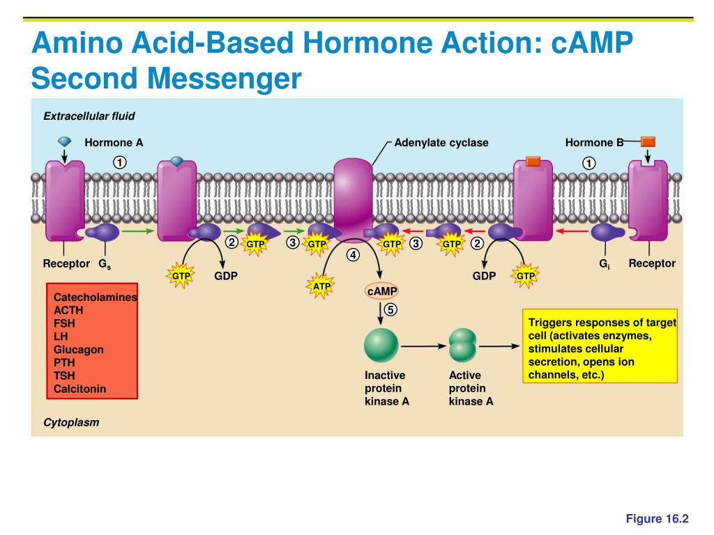
This is one of the most common signaling pathways in the body. It is used by hormones that are critical to the menstrual cycle, including LH and FSH. The pathway is centered on the second messenger cyclic AMP (cAMP).
The process involves these steps:
- A hormone binds to a GPCR linked to a stimulatory G protein (Gs).
- The activated Gs-alpha subunit binds to and activates an enzyme called adenylyl cyclase.
- Adenylyl cyclase converts ATP (the cell's main energy currency) into cAMP.
- cAMP molecules then bind to and activate another enzyme called Protein Kinase A (PKA).
- Active PKA is a kinase, meaning its job is to add phosphate groups to other proteins (phosphorylation). This phosphorylation acts like a switch, altering the activity of various target proteins and enzymes within the cell, leading to the final cellular response (e.g., increased hormone synthesis, opening ion channels).
The image below illustrates this cascade, including an opposing pathway involving an inhibitory G protein (Gi), which blocks adenylyl cyclase to turn the signal down.
Let's watch a detailed walkthrough of this pathway.
Endocrinology | Receptor Pathways
This segment of the Ninja Nerd video provides a fantastic, step-by-step animation of the G-stimulatory (Gs) protein pathway.
Watch from 05:05 to 13:58. Pay close attention to each step: hormone binding, G protein activation (GDP/GTP swap), adenylyl cyclase activation, cAMP production, and PKA activation. Note the wide variety of cellular effects PKA can have at the end.
To turn the signal off, an enzyme called phosphodiesterase (PDE) breaks down cAMP, ensuring the cell's response is transient and tightly controlled.
4. The IP3/DAG Second Messenger Pathway
Another crucial pathway involves a different G protein, Gq, and two different second messengers. This pathway is essential for the action of Gonadotropin-Releasing Hormone (GnRH), the master regulator of the reproductive axis.
The steps are as follows:
- A hormone (like GnRH) binds to a GPCR linked to a Gq protein.
- The activated Gq-alpha subunit binds to and activates an enzyme called Phospholipase C (PLC).
- PLC cleaves a specific membrane phospholipid (PIP₂) into two separate second messengers:
- Diacylglycerol (DAG): This lipid molecule stays in the cell membrane.
- Inositol 1,4,5-trisphosphate (IP₃): This small, water-soluble molecule diffuses into the cytoplasm.
- IP₃ binds to receptors on the endoplasmic reticulum (the cell's internal calcium store), causing channels to open and release a flood of Ca²⁺ ions into the cytoplasm.
- DAG, together with the elevated cytoplasmic Ca²⁺, recruits and activates another enzyme, Protein Kinase C (PKC).
- Active PKC then phosphorylates a different set of target proteins, leading to the cell's response. The released Ca²⁺ also acts as a second messenger itself, binding to proteins like calmodulin to regulate other cellular processes.

The next part of the Ninja Nerd video explains this pathway beautifully.
Endocrinology | Receptor Pathways
Continuing with the Ninja Nerd video, this section explains the Gq protein pathway, detailing the roles of Phospholipase C, IP3, DAG, and calcium release.
Watch from 13:58 to 22:31. Follow how Gq activation leads to the dual signaling branches of IP3 (calcium release) and DAG (PKC activation).
Test your understanding!
A researcher is studying pituitary cells that secrete LH. These cells are stimulated by GnRH, which uses the IP3/DAG pathway. If the researcher adds a drug that specifically blocks Phospholipase C (PLC), what would happen to LH secretion when the cells are exposed to GnRH?
Show answer
If PLC is blocked, it cannot cleave PIP₂ into IP₃ and DAG, even if the Gq protein is activated by GnRH binding. Without IP₃, there will be no release of calcium from the endoplasmic reticulum. Without DAG and calcium, Protein Kinase C will not be activated. As a result, the entire downstream signaling cascade is halted, and the pituitary cell will fail to secrete LH in response to GnRH.
To consolidate your understanding of both major GPCR pathways, the following text provides a concise, academic summary.
The StatPearls article, 'Biochemistry, Hormones', provides an excellent written summary of the mechanisms we've just seen. Reading this will help solidify the terminology and sequence of events.
Please read the first paragraph under the 'Mechanism' section, titled 'Cell Surface Receptors'. Focus on the descriptions of the Gs (cAMP pathway) and Gq (PLC, DAG, IP3 pathway) subunits and the hormones they mediate.
5. An Alternative Pathway: Receptor Tyrosine Kinases (RTKs)
While GPCRs are extremely common, another class of receptors is vital for hormones related to growth and metabolism, most importantly insulin. These are Receptor Tyrosine Kinases (RTKs). Unlike GPCRs, which use a separate G protein, RTKs have their own built-in enzyme activity.
The mechanism is different:
- Hormone Binding & Dimerization: The binding of the hormone (e.g., insulin) causes two receptor molecules to move together and pair up, forming a dimer.
- Autophosphorylation: Dimerization activates the intrinsic kinase function of the receptors. Each receptor in the dimer adds phosphate groups to tyrosine amino acids on its partner. This is called autophosphorylation or cross-phosphorylation.
- Recruitment of Signaling Proteins: The newly phosphorylated tyrosines act as docking sites for various intracellular "adapter proteins" (like Insulin Receptor Substrate, or IRS).
- Downstream Cascade: These adapter proteins, once docked and activated, initiate a complex cascade of further protein phosphorylations inside the cell, ultimately leading to insulin's effects, such as promoting glucose uptake and storage.
The Dirty Medicine channel offers a very clear and concise explanation of all these pathways, including RTKs.
Signal Transduction Pathways (G-Protein, Receptor Tyrosine Kinase, cGMP)
The video 'Signal Transduction Pathways' provides a rapid and high-yield overview of all the pathways we are discussing. It is particularly clear on the RTK mechanism.
Please watch from 02:13 to 14:46. This covers: A review of the G-protein pathways (Gs, Gi, Gq). A list of which hormones use the cAMP vs. IP3 pathways (very relevant!). A clear explanation of the Receptor Tyrosine Kinase pathway. A list of hormones, including insulin, that use the RTK pathway.
Conclusion
Today, we've unraveled the elegant strategies that water-soluble hormones use to communicate with their target cells. Unable to enter directly, they bind to surface receptors and trigger internal chain reactions managed by second messengers. This system allows for both rapid signaling and tremendous amplification.
Key Takeaways:
- Peptide and amine hormones are typically water-soluble and act via cell-surface receptors.
- The hormone is the first messenger; its binding activates intracellular second messengers.
- G Protein-Coupled Receptors (GPCRs) are a major class of receptors that use a G protein intermediate.
- The cAMP pathway (via Gs) involves adenylyl cyclase, cAMP, and Protein Kinase A. It is used by LH and FSH.
- The IP3/DAG pathway (via Gq) involves Phospholipase C, IP3, DAG, and Ca²⁺, activating Protein Kinase C. It is used by GnRH.
- Receptor Tyrosine Kinases (RTKs) are another class of receptors with intrinsic enzyme activity, used by insulin. They work via dimerization and autophosphorylation.
Preview of the Next Lesson:
We have now covered the two major classes of hormone action: direct gene regulation by lipid-soluble hormones and second messenger cascades by water-soluble hormones. In the next lesson, we will take a step back to look at the bigger picture, exploring the general principles of how hormones are made (synthesis), released (secretion), and carried through the body (transport).