Hello! Welcome to the fourth lesson in our module on insulin and glucose metabolism.
Introduction
In our last lesson, we established insulin's role as the master anabolic hormone, directing your cells to take up glucose from the blood and store it as glycogen or fat. We saw how insulin acts as the "key" that unlocks the cell's metabolic machinery.
Today, we address a pivotal question: What happens when the locks become rusty and the key no longer works efficiently? This phenomenon is called insulin resistance, a central concept in the pathophysiology of PCOS. This lesson will focus on our learning outcome: Define insulin resistance and explain its key molecular mechanisms. We will move from the what of insulin's action to the why of its failure, laying a critical foundation for understanding the hormonal imbalances in PCOS.
1. Defining Insulin Resistance
At its core, the definition of insulin resistance is straightforward.
Insulin Resistance: Background, Pathophysiology, Etiology
Let's begin with a formal definition from the medical resource Medscape. This will provide a clear and concise starting point for our discussion.
Please read the 'Background' section of this article. Focus on the first sentence, which provides the essential definition of insulin resistance.
As the article states, insulin resistance is a state where a given concentration of insulin produces a less-than-expected biological effect.
Imagine you're trying to talk to someone across a quiet room; a normal speaking voice works perfectly. Now, imagine the room is filled with loud music; you have to shout to be heard. Insulin resistance is the cellular equivalent of that noisy room. The target tissues (muscle, fat, liver) become "deaf" to insulin's signal. To compensate, the pancreas has to "shout" by secreting much higher amounts of insulin to get the same job done—a state known as hyperinsulinemia. We will explore this compensatory mechanism in our next lesson.
2. A Framework for Understanding the Causes
The failure in insulin signaling can occur at various points along the pathway, from the hormone itself to the final intracellular actions. We can categorize these causes into three main groups.
Insulin Resistance: Background, Pathophysiology, Etiology
The Medscape article provides a useful framework for classifying the causes of insulin resistance. Understanding this classification will help organize the complex molecular details.
Please read the section titled 'Etiology,' focusing on the sub-section 'Classification of prereceptor, receptor, and postreceptor causes.' This will introduce the three main categories of defects.
To summarize the framework:
- Pre-receptor causes: The problem is with the insulin molecule itself (e.g., genetic mutations) or its ability to reach the receptor (e.g., anti-insulin antibodies). These are relatively rare.
- Receptor causes: The problem is with the insulin receptor on the cell surface. There might be too few receptors, or they might be dysfunctional due to mutations or blocking antibodies.
- Post-receptor causes: The problem lies downstream of the receptor, within the intracellular signaling cascade (IRS proteins, PI3K, Akt, etc.). This is the most common cause of the insulin resistance seen in obesity, type 2 diabetes, and PCOS.
Given its importance, we will now focus entirely on the post-receptor mechanisms.
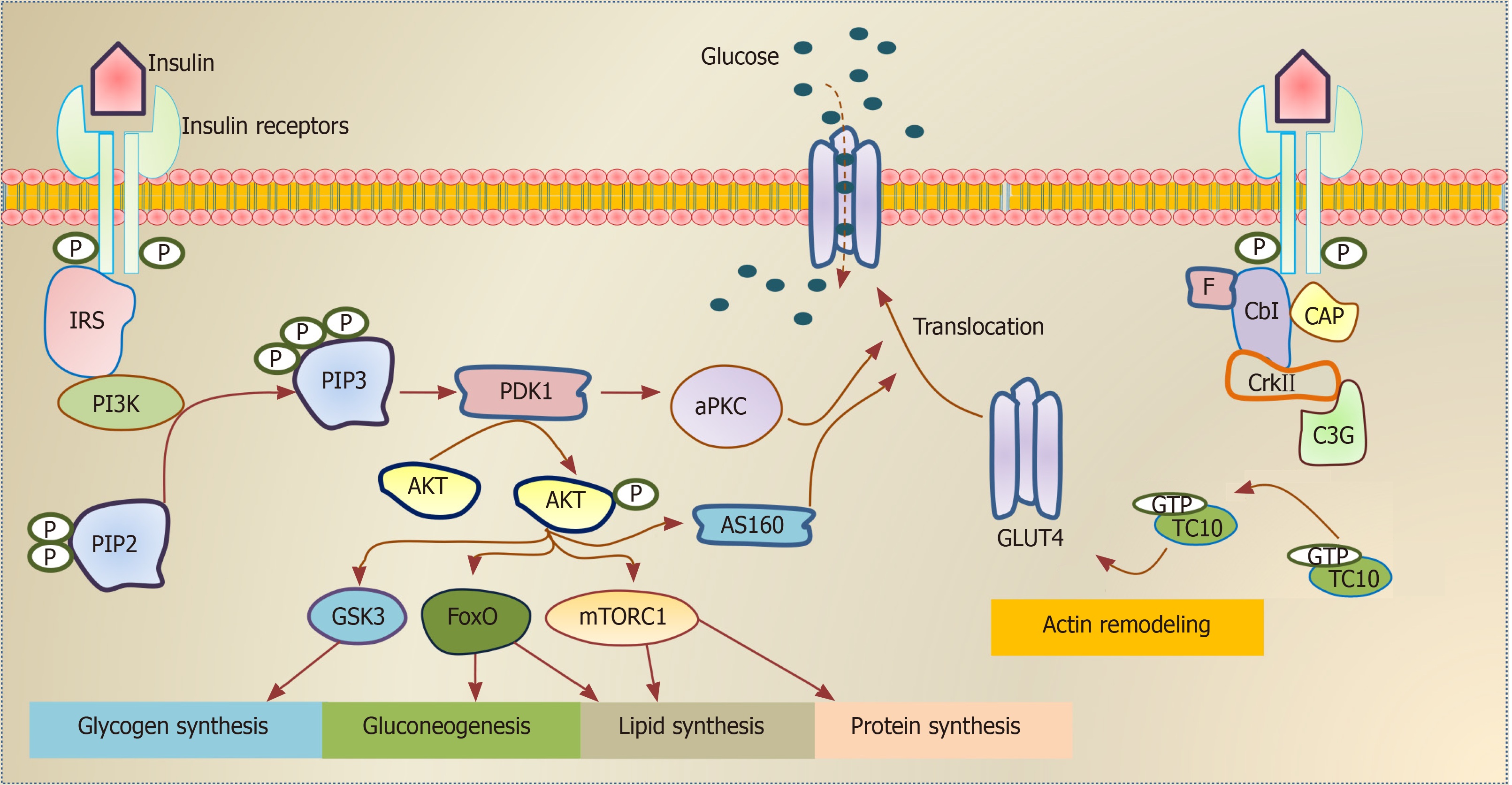
3. The Molecular Mechanisms of Post-Receptor Insulin Resistance
What exactly goes wrong inside the cell to disrupt this finely tuned pathway? While several factors are involved, a leading and well-supported theory points to the accumulation of specific lipid molecules inside the cell.
Dr. Gerald Shulman's research provides a compelling narrative of how scientists identified the precise molecular breakdown.
Step 1: Pinpointing the Defect
The first step was to determine which part of glucose metabolism was failing in insulin-resistant individuals. Was it glucose transport into the cell, or its conversion to glycogen?
In this segment from a lecture at the University of Miami, Dr. Shulman explains how his team used advanced imaging techniques (NMR spectroscopy) to non-invasively 'see' inside the muscle cells of diabetic patients and identify the rate-limiting step in their insulin resistance.
Please watch from 07:49 to 18:13. The key takeaway is that in insulin-resistant individuals, both intracellular glucose and glucose-6-phosphate levels are low, which strongly implicates a defect in glucose transport (i.e., getting glucose into the cell via GLUT4) as the primary bottleneck.
This discovery was crucial: the problem wasn't the cell's ability to use glucose once inside, but its ability to get it inside in the first place. The GLUT4 gates were not opening properly in response to insulin.
Step 2: Unmasking the Culprit—Intracellular Lipids
The next question was, what prevents the GLUT4 gates from opening? The research pointed toward excess fat, not just around the cells, but inside them.
The central mechanism involves these key players:
- Free Fatty Acids (FFAs): When there is an excess of energy (from diet or released from fat stores), the concentration of FFAs inside muscle and liver cells increases.
- Diacylglycerol (DAG): This FFA excess leads to the accumulation of a specific lipid intermediate called diacylglycerol. Think of DAG as a byproduct of an overwhelmed fat-processing factory.
- Protein Kinase C (PKC): DAG acts as a potent activator for a family of enzymes called Protein Kinase C.
Once activated by DAG, PKC becomes the saboteur of the insulin signaling pathway. It interferes with the very first step after the insulin receptor.
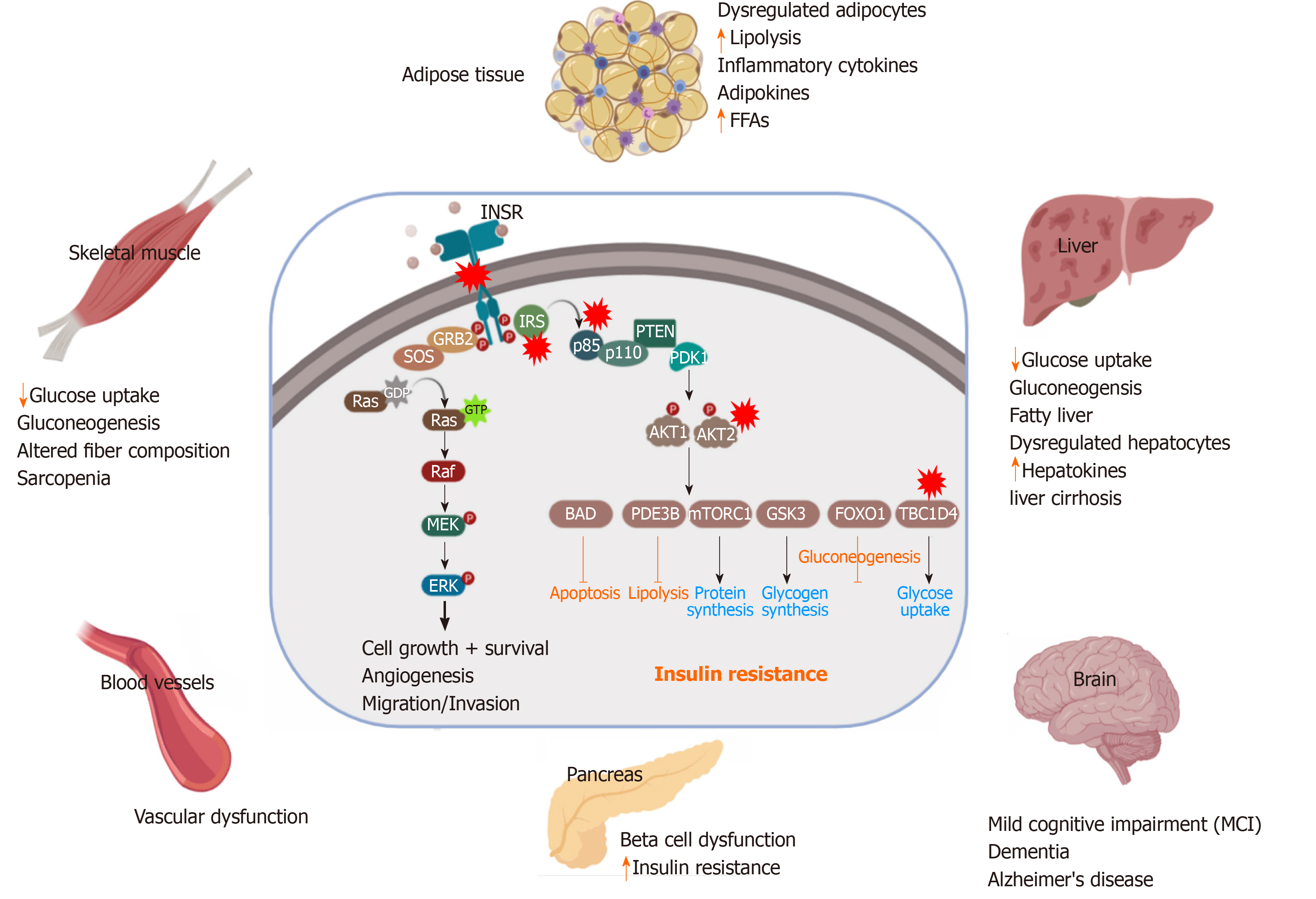
Here is the critical chain of events:
- Activated PKC phosphorylates the Insulin Receptor Substrate (IRS-1).
- Crucially, PKC adds a phosphate group to a serine or threonine amino acid on IRS-1.
- This "wrong" phosphorylation prevents the insulin receptor from performing the "correct" phosphorylation on a tyrosine amino acid of IRS-1.
- Without proper tyrosine phosphorylation, IRS-1 cannot activate the next enzyme in the chain, PI3-Kinase.
- The entire downstream cascade (including Akt activation and GLUT4 translocation) is blocked.
The insulin key is in the lock, but the internal mechanism is jammed.
This next video segment explains this entire process, unifying the link between fat, DAG, PKC, and the ultimate failure of insulin signaling.
Let's return to Dr. Shulman's lecture. He summarizes this 'lipid-induced' insulin resistance mechanism, explaining how diacylglycerol (DAG) accumulation activates PKC, which in turn blocks the insulin signaling cascade.
Please watch from 26:39 to 31:54. Focus on his diagram and explanation of how DAG activates PKC, leading to the inhibition of the insulin receptor's ability to signal through PI3-Kinase. This is the core molecular explanation for the most common form of insulin resistance.
Test your understanding!
A hypothetical drug is developed that specifically prevents diacylglycerol (DAG) from binding to and activating Protein Kinase C (PKC) in muscle cells. Based on the mechanism we just learned, what effect would you predict this drug would have on a person with obesity-related insulin resistance?
Show answer
This drug would likely improve their insulin sensitivity. Even if their muscle cells have high levels of DAG due to excess fat, the drug would prevent DAG from activating PKC. Without activated PKC, the "sabotage" of the IRS-1 protein would not occur. This would allow the insulin signaling pathway to function more normally, leading to better GLUT4 translocation and improved glucose uptake by the muscle cells.
Other Post-Receptor Defects
While the lipid-induced mechanism is a major contributor, it's also important to know that inherited genetic defects in the signaling proteins themselves can cause insulin resistance. These are less common but highlight the importance of each link in the signaling chain.
Insulin signaling and its application
To broaden our understanding, this review article from 'Frontiers in Endocrinology' describes several genetic mutations in the post-receptor pathway that are known to cause insulin resistance.
Please read sections 3 ('Insulin resistance mechanism'), 3.1 ('Substrates of insulin signaling'), and 3.2 ('Negative modulators of insulin signaling'). You don't need to memorize the specific mutations; the goal is to appreciate that defects in genes for IRS-1, PI3K, and AKT can directly impair the pathway and lead to insulin resistance.
Conclusion
Today, we've defined insulin resistance as a state of cellular deafness to insulin's signal and delved deep into the molecular "why." We've moved from a simple concept to a detailed mechanistic understanding, which is crucial for grasping the origins of PCOS.
Key Takeaways:
- Insulin resistance is a condition where a normal amount of insulin produces a sub-par biological response, forcing the pancreas to secrete more insulin to compensate.
- The causes can be classified as pre-receptor, receptor, or post-receptor, with post-receptor defects being the most common in conditions like obesity and PCOS.
- A primary molecular mechanism of post-receptor resistance is lipid-induced dysfunction.
- Excess intracellular fatty acids lead to the accumulation of diacylglycerol (DAG), which activates Protein Kinase C (PKC).
- Activated PKC disrupts insulin signaling by incorrectly phosphorylating IRS-1, preventing the activation of PI3-Kinase and blocking downstream effects like GLUT4-mediated glucose uptake.
Preview of the Next Lesson:
We've established that in a state of insulin resistance, the pancreas "shouts" louder by secreting more insulin. What are the consequences of this chronically elevated insulin level, or compensatory hyperinsulinemia? In our next lesson, we will explore this crucial consequence and see how it becomes a major driver of the metabolic and reproductive problems seen in PCOS.